. 2004 Jul;10(7):1034-46.
doi: 10.1261/rna.7300804.

Pseudouridine-guide RNAs and other Cbf5p-associated RNAs in Euglena gracilis

Affiliations

Pseudouridine-guide RNAs and other Cbf5p-associated RNAs in Euglena gracilis

Anthony G Russell et al. RNA. 2004 Jul.

Abstract

In eukaryotes, box H/ACA small nucleolar RNAs (snoRNAs) guide sites of pseudouridine (Psi) formation in rRNA. These snoRNAs reside in RNP complexes containing the putative Psi synthase, Cbf5p. In this study we have identified Cbf5p-associated RNAs in Euglena gracilis, an early diverging eukaryote, by immunoprecipitating Cbf5p-containing complexes from cellular extracts. We characterized one box H/ACA-like RNA which, however, does not appear to guide Psi formation in rRNA. We also identified four single Psi-guide box AGA RNAs. We determined target sites for these putative Psi-guide RNAs and confirmed that the predicted Psi modifications do, in fact, occur at these positions in Euglena rRNA. The Cbf5p-associated snoRNAs appear to be encoded by multicopy genes, some of which are clustered in the genome together with methylation-guide snoRNA genes. These modification-guide snoRNAs and snoRNA genes are the first ones to be reported in euglenid protists, the evolutionary sister group to the kinetoplastid protozoa. Unexpectedly, we also found and have partially characterized a selenocysteine tRNA homolog in the anti-Cbf5p-immunoprecipitated sample.


Figures
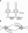
FIGURE 1.
FIGURE 1.
Structure and function of the two classes of modification-guide RNA. (A) A eukaryotic box H/ACA RNA. The two extended helices are interrupted by internal bulge regions that base pair to sequences flanking the unpaired uridine in the substrate RNA, specifying its conversion to Ψ. In this example, the box H/ACA guide RNA is depicted as specifying two sites of pseudouridine formation in the same target RNA species. (B) A eukaryotic box C/D RNA. The conserved sequence motifs are shown boxed and are juxtaposed to form part of the K-turn structural element (Watkins et al. 2000; Klein et al. 2001). Helical elements found in some eukaryotic box C/D RNAs are shown. The region immediately 5′ to the D and/or D′ box pairs with the target RNA. The nucleotide in the target RNA that is O2′-methylated, indicated by the black circle, is paired to the residue exactly 5 nt from the D or D′ box.
FIGURE 2.
FIGURE 2.
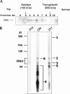
Purification and visualization of E. gracilis Cbf5p-associated RNAs. E. gracilis crude cell extracts were fractionated on glycerol gradients, and Cbf5p-containing complexes were immunoprecipi-tated. (A) Western blot analysis of glycerol gradient fractions. Protein components from equivalent volumes of each gradient fraction were resolved by SDS-PAGE and probed with αCbf5p. Protein size markers were run on a parallel gradient, and their migration positions are indicated at the top. (B) Visualization of immunoprecipitated RNAs. αCbf5p RNAs immunoprecipitated from the concentrated glycerol gradient fraction were radioactively 3′ end-labeled and resolved by electrophoresis in an 8% denaturing polyacrylamide gel. The position of migration of size markers is indicated at the left, as is the position of migration of tRNAs. Immunoprecipitation was performed overnight (“ON”) or for 1 h (“1hr”). Immunoprecipitation with pre-immune serum (“Pre”) was performed for 1 h. RNA species for which sequencing reactions were performed are labeled as bands 1–4 and 6. Distinct RNA species were also isolated from region 5 of the overnight immunoprecipitation sample and sequenced.
FIGURE 3.
FIGURE 3.
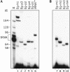
Detection by RT primer extension of small RNAs in E. gracilis total RNA. The name of each RNA analyzed is indicated at the top of each lane. (A) Detection of RNAs found associated with E. gracilis Cbf5p. Lane M is a size marker generated by 3′ end-labeling E. gracilis total RNA. Sizes (in nt) of some of the LSU rRNA species and the position of migration of tRNAs are indicated on the left. Because the primers used contained an 11-nt 5′ anchor sequence and the complementary region was not always exactly at the 3′ end (see Figs. 4 ▶, 6 ▶, 7 ▶), the primer extension products are expected to be longer than the mature RNA by the following number of nt: Eg-h1, +9; Eg-p1, +11; Eg-p2, +1; Eg-p3, +8; Eg-p4, +10; tRNASec, +7. The band marked with * in lane 1 was also characterized (see text). (B) Detection of box C/D RNAs predicted from genomic sequences. These primers also contained the 11-nt anchor, and in each case the complementarity started at the 3′ end of the box-D sequence.
FIGURE 4.
FIGURE 4.
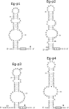
Secondary structures of E. gracilis box AGA modification-guide RNAs. In the secondary structures the conserved AGA motif is boxed. Sites of sequence heterogeneity in the RNAs are indicated in parentheses. For each RNA, the arrow indicates the 3′-most nucleotide that anneals to the RT primer used for the experiments of Figure 3 ▶. GenBank accession nos. are AY572245, AY572242, AY572244, and AY572243 for the Eg-p1 to Eg-p4 RNAs, respectively.
FIGURE 5.
FIGURE 5.
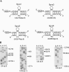
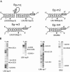
Target sites of pseudouridine formation in rRNA guided by E. gracilis box AGA RNAs. (A) Bipartite base-pairing interactions of the box AGA guide RNAs (top strand) to their predicted rRNA target site (bottom strand). The hairpin structural element in the guide RNA that interrupts the base-pairing interaction is shown schematically. Sites of pseudouridine formation are indicated by “Ψ”. Numbering of nucleotides in the mature LSU rRNA begins at the 5′ end of the 5.8S rRNA as position 1. The LSU species in which a Ψ target site resides is indicated in parentheses; that is, LSU2754 (sp. 8) is nucleotide 2754, located in LSU species 8. In each case, the position marked by * is identically marked in the corresponding autoradiogram shown in (B). (B) Autoradiograms of chemical sequencing gels of 3′ end-labeled E. gracilis 5.8S rRNA and an internal fragment of LSU rRNA species 8. Blanks in the U-specific ladders indicate the positions of Ψ residues, with those predicted from guide RNA sequences denoted by filled triangles.
FIGURE 6.
FIGURE 6.
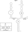
Structure of an E. gracilis box H/ACA RNA (GenBank accession no. AY572240). The conserved H and ACA motifs are shown boxed. The inset depicts a potential base-pairing interaction between stem structure II and LSU species 8. The dashed line highlights nucleotides absent from the smaller RT primer extension product generated from this RNA (see text). The arrow indicates the 3′-most nucleotide that anneals to the RT primer.
FIGURE 7.
FIGURE 7.
E. gracilis selenocysteine tRNASec. (A) Cloverleaf secondary structural model of E. gracilis tRNASec (GenBank accession no. AY572241) in the 9/4 representation. The CU dinucleotide sequence, indicated by * in (B), is bolded. The gray box highlights the residues of the TΨC stem that are discussed in the text. The arrow marks the 3′-most nucleotide that anneals to the RT primer. (B) Multiple sequence alignment of some eukaryotic Sec tRNAs. RNA sequences shown are inferred from the reported gene sequences. Identical positions have a black background; conserved positions have a gray background. The most highly conserved region, the anticodon stem and loop, is delineated. * indicates two nucleotides uniquely variant in the E. gracilis sequence. Sources of the sequences are: Eg, Euglena gracilis (this study); Cr, Chlamydomonas reinhardtii (Rao et al. 2003); Dm, Drosophila melanogaster (Lowe and Eddy 1997; Genomic tRNA database [GtRDB]); Ce, Caenorhabditis elegans (Lowe and Eddy 1997; GtRDB).
FIGURE 8.
FIGURE 8.
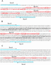
Organization of E. gracilis modification-guide RNA genes. Genomic sequences between copies of Ψ-guide RNA genes for Eg-p2, accession no. AY572246 (A), Eg-p3, accession no. AY572247 (B) and Eg-p1, accession no. AY572248 (C). Capitalized sequence (shown in blue) marks the 5′ and 3′ end sequences of the box AGA RNA genes. The arrows indicate sequences to which the primers anneal. In the intergenic sequences (set in lowercase), regions encoding box C/D RNAs are shown in red and bolded, with the characteristic C- and D-box motifs shown boxed. Regions with extensive complementarity to E. gracilis rRNA are underlined. In B, copies of an imperfect direct repeat are shown in bold and italics.
FIGURE 9.
FIGURE 9.
Target sites for O2′-methylation in rRNA guided by E. gracilis box C/D RNAs. (A) Predicted base-pairing interactions between the box C/D RNAs and sites of E. gracilis rRNA O2′-methylation. The relevant portion of the box C/D guide RNA (top strand) is shown paired to the rRNA (bottom strand), and the nucleotide position of modification is underscored and indicated with a filled circle. Numbering of rRNA positions is as described in Figure 5 ▶. In each case, the position marked by * is identically marked in the corresponding autoradiogram shown in B. (B) Autoradiograms of sequencing gels containing RNase T1 (G-specific) and alkali (alk) ladders of 3′ end-labeled E. gracilis LSU rRNA species 6, 7, 9 and an internal fragment of SSU rRNA. O2′-methyl residues are blank in the alkali ladders, with those predicted from guide RNA sequences indicated by filled triangles.

Similar articles

Cited by

References

    1. Atzorn, V., Fragapane, P., and Kiss, T. 2004. U17/snR30 is a ubiquitous snoRNA with two conserved sequence motifs essential for 18S rRNA production. Mol. Cell. Biol. 24: 1769–1778. - PMC - PubMed
    1. Bachellerie, J.-P., Cavaillé, J., and Hüttenhofer, A. 2002. The expanding snoRNA world. Biochimie 84: 775–790. - PubMed
    1. Bakin, A., Lane, B.G., and Ofengand, J. 1994. Clustering of pseudouridine residues around the peptidyltransferase center of yeast cytoplasmic and mitochondrial ribosomes. Biochemistry 33: 13475–13483. - PubMed
    1. Balakin, AG., Smith, L., and Fournier, M.J. 1996. The RNA world of the nucleolus: Two major families of small RNAs defined by different box elements with related functions. Cell 86: 823–834. - PubMed
    1. Barneche, F., Gaspin, C., Guyot R., and Echeverria, M. 2001. Identification of 66 box C/D snoRNAs in Arabidopsis thaliana: Extensive gene duplications generated multiple isoforms predicting new ribosomal RNA 2′-O-methylation sites. J. Mol. Biol. 311: 57–73. - PubMed

Publication types

LinkOut - more resources