. 2004 Jul;10(7):1097-107.
doi: 10.1261/rna.7140604.

A horizontally acquired group II intron in the chloroplast psbA gene of a psychrophilic Chlamydomonas: in vitro self-splicing and genetic evidence for maturase activity

Affiliations

A horizontally acquired group II intron in the chloroplast psbA gene of a psychrophilic Chlamydomonas: in vitro self-splicing and genetic evidence for maturase activity

Obed W Odom et al. RNA. 2004 Jul.

Abstract

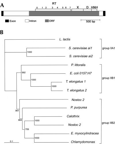
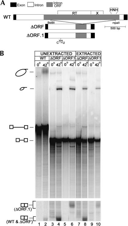
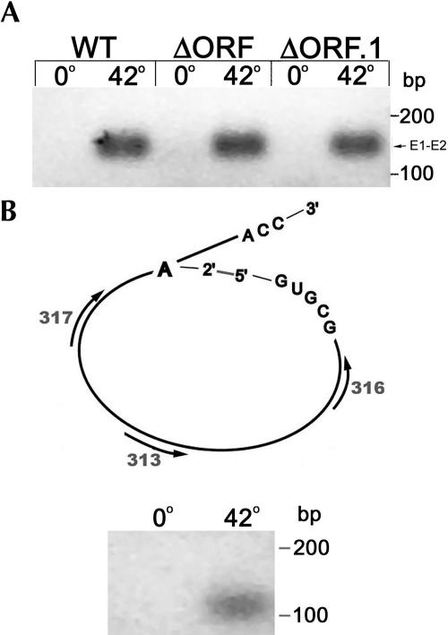
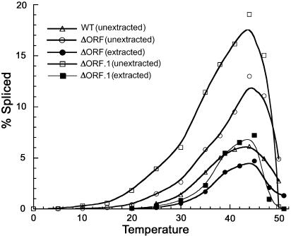
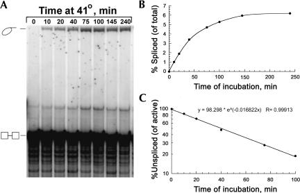
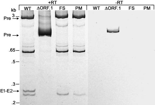
The majority of known group II introns are from chloroplast genomes, yet the first self-splicing group II intron from a chloroplast gene was reported only recently, from the psbA gene of the euglenoid, Euglena myxocylindracea. Herein, we describe a large (2.6-kb) group II intron from the psbA gene (psbA1) of a psychrophilic Chlamydomonas sp. from Antarctica that self-splices accurately in vitro. Remarkably, this intron, which also encodes an ORF with putative reverse transcriptase, maturase, and endonuclease domains, is in the same location, and is related to the E. myxocylindracea intron, as well as to group IIB2 introns from cyanobacteria. In vitro self-splicing of Chs.psbA1 occurred via a lariat, and required Mg(2+) (>12 mM) and NH(4)(+). Self-splicing was improved by deleting most of the ORF and by using pre-RNAs directly from transcription reactions, suggestive of a role for folding during transcription. Self-splicing of Chs.psbA1 pre-RNAs showed temperature optima of ~44 degrees C, but with a broad shoulder on the low side of the peak; splicing was nearly absent at 50 degrees C, indicative of thermolability. Splicing of wild-type Chs.psbA1 also occurred in Escherichia coli, but not when the ORF was disrupted by mutations, providing genetic evidence that it has maturase activity. This work provides the first description of a ribozyme from a psychrophilic organism. It also appears to provide a second instance of interkingdom horizontal transfer of this group IIB2 intron (or a close relative) from cyanobacteria to chloroplasts.


Figures

FIGURE 1.
FIGURE 1.
Light microscope image of Chlamydomonas sp. CCMP-1619. The cells were grown in liquid medium at 4°C, and the image was captured with a digital camera at a total magnification of 600×. The scale bar is 10 μm.
FIGURE 2.
FIGURE 2.
Proposed secondary structure of the 2572-bp Chs.psbA1 intron. Intron nucleotides are in uppercase and numbered from the 5′ end; exon nucleotides are in lowercase. 5′SS and 3′SS are the splice sites. The major domains are labeled with Roman numerals (I–VI) and subdomains of domain I as A, B, C (C1 and C2), and D (D′, D‴, D2a, D2b, D3). Tertiary interactions are labeled with Greek letters (α-α′, etc.). EBS and IBS refer to exon and intron binding sites, respectively. The Shine–Dalgarno (SD) sequence and start codon (AUG) of the ORF in domain IV are boxed, and the branch-site A in domain VI is circled. An alternative structure of the unusual insertion in domain D (nt 275–310) is proposed for the ΔORF.1 mutant (boxed sequence above the wild-type structure).
FIGURE 3.
FIGURE 3.
Domain structure and phylogenetic analysis of the Chlamydomonas sp. (CCMP-1619) Chs.psbA1 intron ORF. (A) Diagram of the intron and ORF subdomains. (RT) reverse transcriptase; (X) maturase; (D) DNA-binding; (HNH) endonuclease. (B) Phylogenetic analysis of the ORF. The neighbor-joining tree was generated with clustal X using amino acid sequences of selected group II intron ORFs. The scale bar is 0.1 substitutions per site. The bootstrap values are out of 1000 possible trees, and the L. lactis protein was the out-group. The ORFs contained all of the domains indicated in A. The ORF sequences and their accession numbers are: Calothrix, Calothrix sp. (X71404.1:1034–2788); Chlamydomonas, Chlamydomonas sp. CCMP-1619 chloroplast psbA intron; E. coli, Escherichia coli 0157:H7 (AF074613.1:58,849–60,573); E. myxocylindracea, Euglena myxocylindracea chloroplast psbA intron (AY290861); L. lactis, LtrA of Lactococcus lactis (AAB06503.1); Nostoc 1, Nostoc sp. PCC 7120 (AP003604.1:45,989–47,791); Nostoc 2, Nostoc sp. PCC 7120 (AP003599.1:30,840–32,459); P. purpurea, Porphyra purpurea mitochondrial rRNA intron 1 (AF114794.1:2909–4543); P. littoralis, Pylaiella littoralis mitochondrial rRNA intron 2 (AJ277126.1:4664–6370); S. cerevisiae ai1, Saccharomyces cerevisiae mitochondrial COXI intron ai1 (NP_009310.1); S. cerevisiae ai2, Saccharomyces cerevisiae mitochondrial COXI intron ai2 (NP_009309.1); T. elongatus 1, Thermo-synechococcus elongatus BP-1 (AP005369.1:28791–30,485); T. elongates 2, Thermosynechococcus elongatus BP-1 (AP005371.1:25,718–27,406).
FIGURE 4.
FIGURE 4.
In vitro self-splicing of 32P-labeled Chs.psbA1 pre-RNAs. (A) Maps of the pre-RNAs. Wild-type and ΔORF pre-RNAs have the same 5′ (67 nt) and 3′ (92 nt) exons, whereas the 5′ and 3′ exons of ΔORF.1 have an additional 5 nt. The wild-type intron is 2572 nt, and 997 nt in the ΔORF and ΔORF.1 pre-RNAs. The nucleotide substitution in the ΔORF.1 core is indicated. See Figure 3 ▶ for explanation of ORF domains. (B) Gel analysis of splicing reactions with 32P-labeled pre-RNAs. The reactions were incubated at 0°C or 42°C for 90 min and then analyzed on a denaturing polyacrylamide gel. “Unextracted” RNAs were taken from transcription reactions and used for splicing without treatment; “extracted” RNAs were extracted with chloroform and ethanol precipitated before splicing. The location of excised lariat, precursor, and spliced-exon RNAs are indicated to the left.
FIGURE 5.
FIGURE 5.
RT-PCR analysis of Chs.psbA1 self-splicing and branching. (A) RT-PCR of spliced-exon RNA. RT-PCR was performed with flanking exon primers on splicing reactions incubated at the indicated temperatures (2 h). The portion of the ethidium-agarose gel containing the 148-bp product (E1–E2) that was eluted and sequenced is shown, and the locations of size markers are indicated. The image was captured with a digital camera and then inverted. (B) RT-PCR analysis of the branch site. (Above) A diagram of the excised intron lariat showing the location of the DNA primers used for the RT-PCR. Reverse transcription (RT) was with primer 313, and then nested PCR was performed with primers 316 and 317. (Below) The wild-type pre-RNA was incubated for self-splicing at the indicated temperatures for 2 h, and then RT-PCR was performed. The ethidium bromide-agarose gel of the RT-PCR product (109 bp) that was eluted and sequenced is shown and the locations of size markers are indicated. The image was captured with a digital camera and then inverted.
FIGURE 6.
FIGURE 6.
Self-splicing of Chs.psbA1 pre-RNAs as a function of temperature. The splicing reactions were performed with 32P-labeled pre-RNAs incubated for 90 min at the indicated temperatures. The RNAs were separated on denaturing polyacrylamide gels and quantified with a phosphorimager. The fraction (in percent) of pre-RNA that spliced was determined from the fraction of intron (in pre-RNA) that was converted to lariat. As indicated, both unextracted and extracted pre-RNAs (see Fig. 4 ▶) were used.
FIGURE 7.
FIGURE 7.
Time-course reaction for self-splicing of a Chs.psbA1 pre-RNA lacking most of the ORF. (A) Autoradiograph of a reaction analyzed by denaturing polyacrylamide gel electrophoresis. The 32P-labeled pre-RNA (ΔORF, extracted) was incubated under standard conditions for the indicated times and temperature. The lariat intron and pre-RNA are indicated. (B) Plot of the fraction of pre-RNA (in percent) that spliced via a lariat over the time course. The gel was quantified using a phosphorimager, and the fraction spliced was determined by estimating the fraction of intron (in pre-RNA) that was converted to lariat. (C) Plot of the decline of active pre-RNA over time. The amount of unspliced pre-RNA that was active (which was 6.3% for this experiment) was determined from B.
FIGURE 8.
FIGURE 8.
ORF-dependent splicing of the Chs.psbA1 intron in E. coli. Expression of the intron constructs in E. coli was induced with IPTG, and RNA was extracted and analyzed by RT-PCR with primers flanking the Chs.psbA1 intron. The reverse transcription step was performed with a T3 promoter primer that anneals just downstream from the psbA exon 2 sequence, and then PCR was performed with the same primer plus one in psbA exon 1. The RT-PCR reactions were either complete (+RT) or lacked the reverse transcriptase (-RT). The E. coli clones contained the following forms of the Chs.psbA1 intron: WT, wild-type; ΔORF.1, ORF-deleted; FS, frame-shift mutation after amino acid 10 of the ORF; and PM, two missense (point) mutations, one in the RT domain and one in the maturase (X) domain. The samples were analyzed by electrophoresis in a 6% polyacrylamide gel (native), which was stained with SYBR Green I and photographed with short-wave UV epi-illumination and a digital camera; the image shown was inverted. The positions and sizes of selected DNA size markers and the identification of RT-PCR products from the unspliced (Pre) and spliced-exon (E1–E2) RNAs are indicated to the left.
FIGURE 9.
FIGURE 9.
Northern blot analysis of psbA transcripts in Chlamydomonas sp. CCMP-1619. CCMP-1619 was grown in continuous light at 8°C (lanes L), and then some cells were shifted to darkness for 3 h (lanes D). RNA was isolated and hybridized with either 32P-labeled exon DNA (Exon Probe) or a Chs.psbA1 intron fragment (Intron Probe). RNA from C. reinhardtii (C.r.) was used as a marker for mature psbA mRNA. C. reinhardtii rRNAs on the methylene blue-stained blot were used as size markers.

Similar articles

Cited by

References

    1. Adamidi, C., Fedorova, O., and Pyle, A.M. 2003. A group II intron inserted into a bacterial heat-shock operon shows autocatalytic activity and unusual thermostability. Biochemistry 42: 3409–3418. - PubMed
    1. Bonen, L. and Vogel, J. 2001. The ins and outs of group II introns. Trends Genet. 17: 322–331. - PubMed
    1. Brown, J.W., Haas, E.S., and Pace, N.R. 1993. Characterization of ribonuclease P RNAs from thermophilic bacteria. Nucleic Acids Res. 21: 671–679. - PMC - PubMed
    1. Bunse, A.A., Nickelsen, J., and Kück, U. 2001. Intron-specific RNA binding proteins in the chloroplast of the green alga Chlamydomonas reinhardtii. Biochim. Biophys. Acta 1519: 46–54. - PubMed
    1. Cannone, J.J., Subramanian, S., Schnare, M.N., Collett, J.R., D’Souza, L.M., Du, Y., Feng, B., Lin, N., Madabusi, L.V., Muller, K.M., et al. 2002. The comparative RNA web (CRW) site: An online database of comparative sequence and structure information for ribosomal, intron, and other RNAs. BioMed Central Bioinform. 3: 2. - PMC - PubMed

Publication types

MeSH terms

LinkOut - more resources