Discrimination between uterine serous papillary carcinomas and ovarian serous papillary tumours by gene expression profiling
- PMID: 15208622
- PMCID: PMC2409747
- DOI: 10.1038/sj.bjc.6601791
Discrimination between uterine serous papillary carcinomas and ovarian serous papillary tumours by gene expression profiling
Abstract
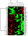
High-grade ovarian serous papillary cancer (OSPC) and uterine serous papillary carcinoma (USPC) represent two histologically similar malignancies characterised by markedly different biological behavior and response to chemotherapy. Understanding the molecular basis of these differences may significantly refine differential diagnosis and management, and may lead to the development of novel, more specific and more effective treatment modalities for OSPC and USPC. We used an oligonucleotide microarray with probe sets complementary to >10 000 human genes to determine whether patterns of gene expression may differentiate OSPC from USPC. Hierarchical cluster analysis of gene expression in OSPC and USPC identified 116 genes that exhibited >two-fold differences (P<0.05) and that readily distinguished OSPC from USPC. Plasminogen activator inhibitor (PAI-2) was the most highly overexpressed gene in OSPC when compared to USPC, while c-erbB2 was the most strikingly overexpressed gene in USPC when compared to OSPC. Overexpression of the c-erbB2 gene and its expression product (i.e., HER-2/neu receptor) was validated by quantitative RT-PCR as well as by flow cytometry on primary USPC and OSPC, respectively. Immunohistochemical staining of serous tumour samples from which primary OSPC and USPC cultures were derived as well as from an independent set of 20 clinical tissue samples (i.e., 10 OSPC and 10 USPC) further confirmed HER-2/neu as a novel molecular diagnostic and therapeutic marker for USPC. Gene expression fingerprints have the potential to predict the anatomical site of tumour origin and readily identify the biologically more aggressive USPC from OSPC. A therapeutic strategy targeting HER-2/neu may be beneficial in patients harbouring chemotherapy-resistant USPC.
Figures


References
-
- Berchuck A, Kamel A, Whitaker R, Kerns B, Olt G, Kinney R, Soper JT, Dodge R, Clarke-Pearson DL, Marks P (1990) Overexpression of HER-2/neu is associated with poor survival in advanced epithelial ovarian cancer. Cancer Res 50: 4087–4091 - PubMed
-
- Budnik LT, Mukhopadhyay AK (2002) Lysophosphatidic acid and its role in reproduction. Biol Reprod 66: 859–865 - PubMed
-
- Carcangiu ML, Chambers JT (1992) Uterine papillary serous carcinoma: a study on 108 cases with emphasis on prognostic significance of associated endometrioid carcinoma, absence of invasion, and concomitant ovarian cancer. Gynecol Oncol 47: 298–305 - PubMed
-
- Carcangiu ML, Chambers JT (1995) Early pathologic stage clear cell carcinoma and uterine papillary serous carcinoma of the endometrium, comparison of clinicopathological features and survival. Int J Gynecol Pathol 14: 30–38 - PubMed
Publication types
MeSH terms
Substances
LinkOut - more resources
Full Text Sources
Other Literature Sources
Medical
Research Materials
Miscellaneous

