. 2004 Aug 12;23(36):6105-14.
doi: 10.1038/sj.onc.1207815.

Mechanisms underlying differential expression of interleukin-8 in breast cancer cells

Affiliations

Mechanisms underlying differential expression of interleukin-8 in breast cancer cells

Ariane Freund et al. Oncogene. .

Abstract

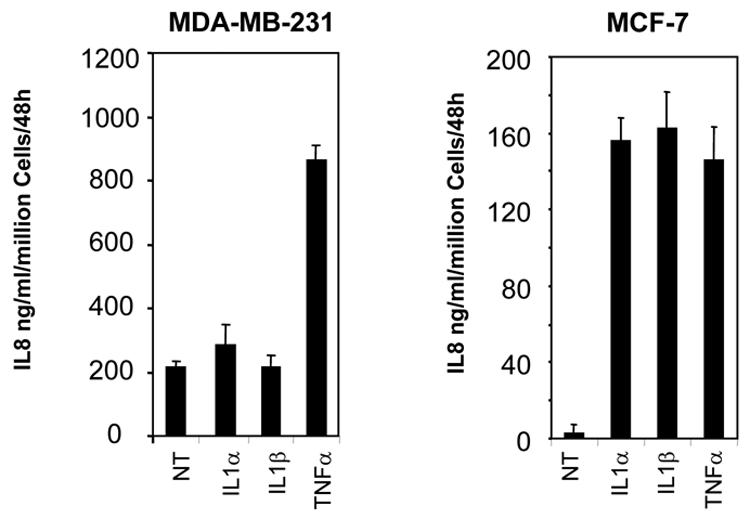
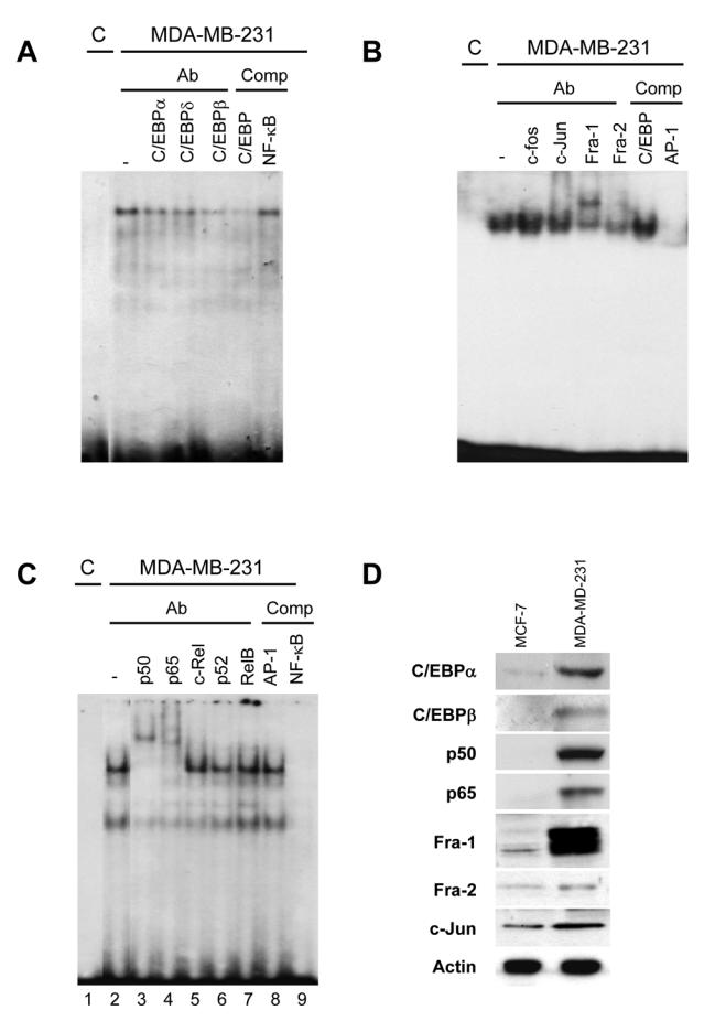
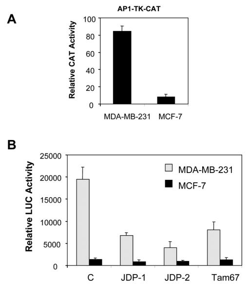
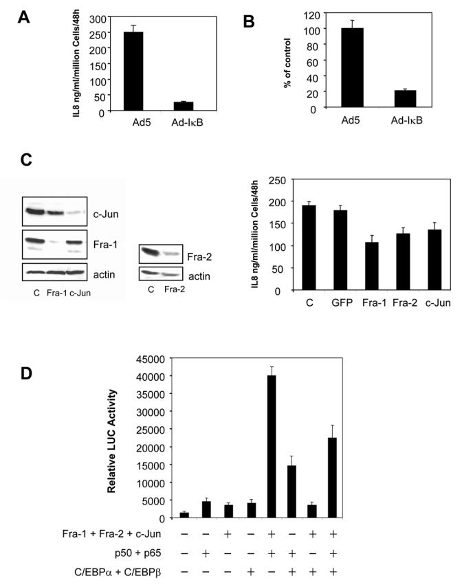
We have recently reported that interleukin-8 (IL-8) expression was inversely correlated to estrogen receptor (ER) status and was overexpressed in invasive breast cancer cells. In the present study, we show that IL-8 overexpression in breast cancer cells involves a higher transcriptional activity of IL-8 gene promoter. Cloning of IL-8 promoter from MDA-MB-231 and MCF-7 cells expressing high and low levels of IL-8, respectively, shows the integrity of the promoter in both cell lines. Deletion and site-directed mutagenesis of the promoter demonstrate that NF-kappaB and AP-1 and to a lesser extent C/EBP binding sites play a crucial role in the control of IL-8 promoter activity in MDA-MB-231 cells. Knockdown of NF-kappaB and AP-1 activities by adenovirus-mediated expression of an NF-kappaB super-repressor and RNA interference, respectively, decreased IL-8 expression in MDA-MB-231 cells. On the contrary, restoration of Fra-1, Fra-2, c-Jun, p50, p65, C/EBPalpha and C/EBPbeta expression levels in MCF-7 cells led to a promoter activity comparable to that observed in MDA-MB-231 cells. Our data constitute the first extensive study of IL-8 gene overexpression in breast cancer cells and suggest that the high expression of IL-8 in invasive cancer cells requires a complex cooperation between NF-kappaB, AP-1 and C/EBP transcription factors.


Figures

Fig. 1
Fig. 1. IL-8 gene is functional in MDA-MB-231 and MCF-7 cells
Cells were treated for 48h with mock (NT), IL-1α (50 ng/ml), IL-1β (250 pg/ml), TNFα (50 ng/ml). Media were then collected to evaluate IL-8 levels by ELISA. Results are expressed as ng IL-8/ml/48h/million cells and represent the mean ± SD of three independent experiments.
Fig. 2
Fig. 2. The differential expression of IL-8 gene in MDA-MB-231 and MCF-7 cells takes place at the transcriptional level
A. IL8 RNA levels were quantified in MDA-MB-231 and MCF-7 cells by quantitative PCR. Results are expressed as arbitrary units corresponding to the ratio of IL-8 levels normalized by rS9 levels (n=3). B. The transcription rates of IL-8 and rS9 genes in MDA-MB-231 and MCF-7 cells were determined by run on assay. A representative experiment is shown here and the transcription rate, expressed in arbitrary units as the ratio of IL-8 signal over rS9 signal, represents the mean ± SD of three independent experiments. C. IL-8 promoter was cloned in sense (xp2-IL8) or anti-sense orientation (xp2-IL8 AS) in xp2 reporter vector. MDA-MB-231 and MCF-7 cells were transfected with the different constructs along with CMV-GAL internal control. Results show relative luciferase activities (n=3) after normalization for β-galactosidase activity. D. Secreted IL-8 levels from MDA-MB-231, MDA-MB-436, CAMA-1 and MCF-7 cells were determined by ELISA (left panel). Results represent the mean ± SD of three independent experiments. The cell lines mentioned above were transfected with xp2 or xp2-IL8 constructs along with CMV-GAL reporter (right panel). Results show fold activities (ratio of xp2-IL8 activity over xp2) after normalization for β-galactosidase activity (n=3).
Fig. 3
Fig. 3. Determination of IL-8 promoter sequences involved in differential regulation of IL-8 gene activity
MDA-MB-231 and MCF-7 cells were transfected with reporter constructs corresponding to the first 1481, 272, 98 or 50 bp of IL-8 promoter. Results show relative luciferase activities (n=3) after normalization for β-galactosidase activity.
Fig. 4
Fig. 4. NF-κB site is crucial for the high activity of IL-8 promoter
A. IL-8 promoter constructs harboring single, double or triple mutations of AP-1, C/EBP or NF-κB sites were transfected in MDA-MB-231 and MCF-7 cells. Results show relative luciferase activities (n=3). B. Binding of nuclear factors to AP-1, C/EBP and NF-κB sites was examined by gel shift assays using 2 μg of nuclear extracts of MDA-MB-231 and MCF-7 cells. The right panel is a control of nuclear extract loading which corresponds to a western blotting of HDAC-1 antibody against MCF-7 and MDA-MB-231 nuclear extracts.
Fig. 5
Fig. 5. Identification of the factors of MDA-MB-231 cells bound to IL-8 promoter
A. Factors bound to C/EBP site of IL-8 promoter in MDA-MB-231 cells were identified by gel shift assay using C/EBPα, C/EBPδ and C/EBPβ antibodies. Competition with a 10 fold excess of cold consensus C/EBP or NF-κB oligonucleotides confirms the specificity of the complexes. B. The identification of AP-1 members of MDA-MB-231 cel extracts bound to AP-1 site was performed by gel shift assay using c-fos, c-Jun, Fra-1 and Fra-2 antibodies. Competition with a 10 fold excess of cold C/EBP or consensus AP-1 oligonucleotides confirms the specificity of the complexes. C. NF-κB proteins present in MDA-MB-231 cells which bound to the NF-κB site were identified by gel shift assay using p50, p65, c-Rel, p52 and RelB antibodies. Oligonucleotides competition were done with a 10 fold excess of cold AP-1 or consensus NF-κB probes. D. Protein levels of Fra-1, Fra-2, c-jun, p50, p65, C/EBPα and C/EBPβ were determined by western blot using 30 μg of MDA-MB-231 and MCF-7 whole cell extracts. The upper band observed on Fra-1 blot corresponds to the phosphorylated form of the protein.
Fig. 6
Fig. 6. AP-1 pathway is important for IL-8 gene activity
A. AP-1 relative activity was assessed in MDA-MB-231 and MCF-7 cells by transfection of an AP-1 reporter. Results show relative CAT activities (n=3) after normalization for β-galactosidase activity. B. The importance of AP-1 pathway in IL-8 promoter activity regulation was evaluated by transfection of 100 ng of empty vector (C) or expression vectors of JDP1, JDP2 and Tam67 AP-1 inhibitors in MDA-MB-231 and MCF-7 cells, along with xp2-IL8 construct. Results show relative luciferase activities (n=3).
Fig. 7
Fig. 7. NF-κB factors are the primary regulators of in IL-8 promoter activity
A. NF-κB relative activity was evaluated in MDA-MB-231 and MCF-7 cells by transfection of a NF-κB reporter. Results show relative luciferase activities (n=3). B. The importance of NF-κB in IL-8 promoter activity regulation was evaluated by infection of MDA-MB-231 and MCF-7 cells with Ad5 and Ad-IκB viruses (MOI 100) and transfection of xp2-IL8 construct. Results show relative luciferase activities (n=3). C. xp2-IL-8, NF-κB and AP-1 reporters were transfected in MDA-MB-231 cells along with CMV-GAL and control CMV5 or CMV-hERα expression vectors. Cells were treated or not with estradiol (10-8M) for 24h. Results show relative luciferase and CAT activities (n=3).
Fig. 8
Fig. 8. NF-κB, AP-1 and C/EBP transcription factors are acting together to control IL-8 gene expression
A. MDA-MB-231 cells were infected at MOI 100 with backbone Ad5 or Ad-IκB viruses. 24h after infection, the medium was collected and assayed for IL-8 levels by ELISA. Results represent the mean ± SD of three independent experiments. B. RNA were collected from the same cells and used to measure IL-8 RNA levels by quantitative PCR. Results are expressed as arbitrary units corresponding to the ratio of IL-8 levels normalized by rS9 levels (n=3). C. Reduction of IL-8 expression using RNA interference against Fra-1, Fra-2 or c-Jun. MDA-MB-231 cells were transfected with siFra-1, siFRa-2, sic-Jun or siGFP (as control). The extent of silencing of was determined by western blot using antibodies against Fra-1, Fra-2, c-Jun or β-actin (left panel). Secreted IL-8 levels were measured by ELISA 48h afer transfection. Results represent the mean ± SD of three independent experiments. D. xp2-IL8 reporter was cotransfected along with 100 ng of expression vectors of Fra-1, Fra-2, c-Jun, p50, p65, C/EBPα or C/EBPβ in MCF-7 cells. Results show relative luciferase activities (n=3).

References

    1. Bharti AC, Aggarwal BB. Biochem Pharmacol. 2002;64:883–8. - PubMed
    1. Brew R, Southern SA, Flanagan BF, McDicken IW, Christmas SE. Eur J Cancer. 1996;32A:2142–7. - PubMed
    1. Brown PH, Alani R, Preis LH, Szabo E, Birrer MJ. Oncogene. 1993;8:877–86. - PubMed
    1. Ferrer FA, Miller LJ, Andrawis RI, Kurtzman SH, Albertsen PC, Laudone VP, Kreutzer DL. Urology. 1998;51:161–7. - PubMed
    1. Freund A, Chauveau C, Brouillet JP, Lucas A, Lacroix M, Licznar A, Vignon F, Lazennec G. Oncogene. 2003;22:256–65. - PMC - PubMed

Publication types