. 2004 Jul;142(6):933-42.
doi: 10.1038/sj.bjp.0705785. Epub 2004 Jun 21.

Irbesartan inhibits human T-lymphocyte activation through downregulation of activator protein-1

Affiliations

Irbesartan inhibits human T-lymphocyte activation through downregulation of activator protein-1

Shu-Meng Cheng et al. Br J Pharmacol. 2004 Jul.

Abstract

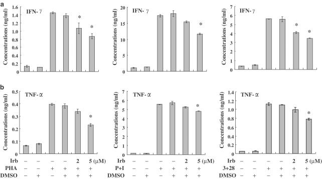
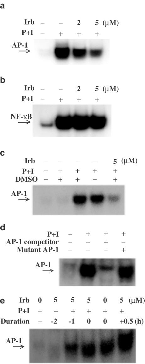
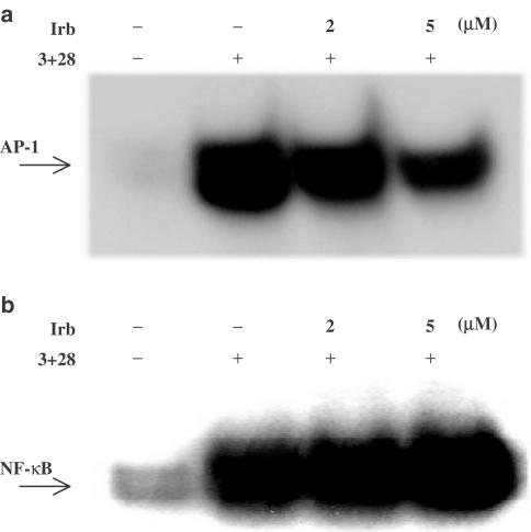
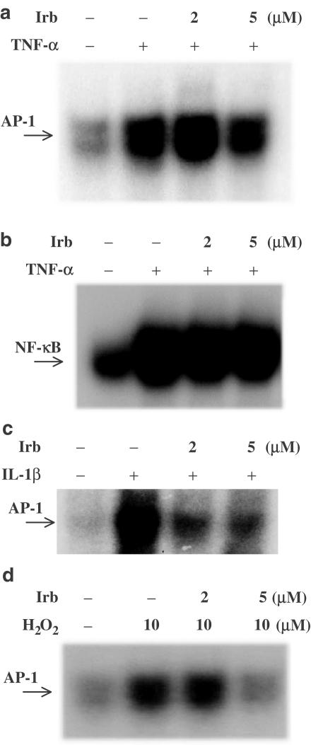
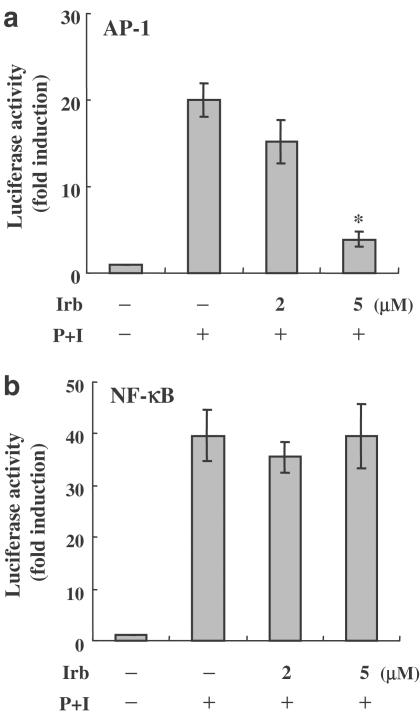
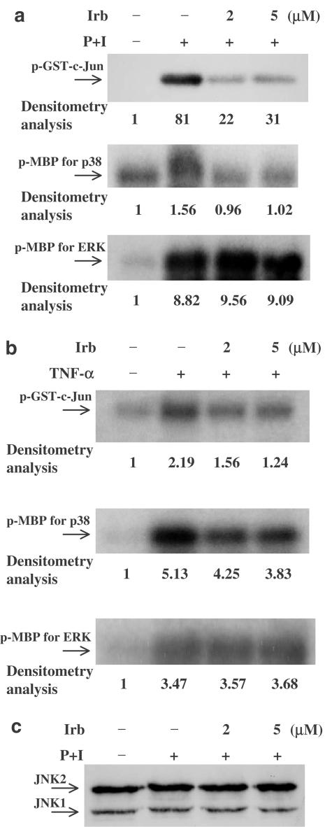
1 Irbesartan is a promising antihypertensive drug with beneficial effects on atherosclerotic processes. In the progression of atherosclerosis, human T-lymphocytes play an important role, but it is not yet known how irbesartan modulates human T-lymphocytes activation. To gain insight into the mechanisms by which irbesartan acts, we investigated its effects on human T-lymphocytes. 2 Primary human T-lymphocytes were isolated from whole blood. Cytokines were determined by ELISA. Activator protein-1 (AP-1) and related protein activities were determined by electrophoretic mobility shift assays, kinase assays, Western blotting and transfection assays. 3 Irbesartan inhibited the production of both tumor necrosis factor-alpha and interferon-gamma by activated T-cells, especially at therapeutic concentrations. Further investigation at the molecular level indicated that the inhibition of activated human T-lymphocytes specifically correlated with the downregulation of AP-1 DNA-binding activity. In the Jurkat T-cell line, irbesartan also inhibited AP-1 transcriptional activity. Finally, we revealed that irbesartan is unique in its ability to inhibit the activation of both c-Jun NH2-terminal protein kinase and p38 MAPK. 4 Our studies show that irbesartan may modulate inflammation-based atherosclerotic diseases through a cell-mediated mechanism involving suppression of human T-lymphocytes activation via downregulation of AP-1 activity.


Figures

Figure 1
Figure 1
Effects of irbesartan (Irb) on cytokine production from activated human T-cells. Human peripheral blood T-cells at 1 × 10⁶ ml−1 were pretreated with various concentrations of irbesartan (2–5 μM) for 2 h and then stimulated with PHA, PMA+ionomycin (P+I) or anti-CD3+anti-CD28 mAb (3+28) for another 24 h. The supernatants were collected for measurements of IFN-γ (a) and TNF-α (b). The data are representative of at least three different donor cells and are shown as means±s.d. * denotes the statistical significance (P-value <0.05) compared with stimulated control cells.
Figure 2
Figure 2
Irbesartan (Irb) blocked AP-1, but not NF-κB DNA-binding activity stimulated by PMA+ionomycin (P+I). Human peripheral blood T-cells at 2 × 10⁶ ml−1 were pretreated with various concentrations of irbesartan for 2 h and then stimulated or not with P+I for 1 h. (a) Irbesartan inhibited AP-1 DNA-binding activity. (b) The activation of NF-κB was not inhibited by irbesartan up to 5 μM. (c) AP-1 activity induced by P+I was not influenced by addition of DMSO at less than 0.05%. (d) The specificity of AP-1 binding activity was also assessed by competition with 100 × unlabeled AP-1. Coincubation of nuclear extracts with 100-fold molar excess of wild-type AP-1 led to inhibition of the retarded AP-1 band. In contrast, nuclear extracts coincubated with mutant-type AP-1 had little effect on the band density. (e) When the cells were pretreated for over 60 min with irbesartan, marked inhibition of AP-1 was observed. Cotreatment or post-stimulus treatment with irbesartan did not inhibit AP-1 activation significantly. Representative data from three different donor cells are shown.
Figure 3
Figure 3
Irbesartan (Irb) blocked AP-1, but not NF-κB DNA-binding activity stimulated by anti-CD3+anti-CD28 mAb. Human peripheral blood T-cells at 2 × 10⁶ ml−1 were pretreated with various concentrations of irbesartan for 2 h and then stimulated or not with anti-CD3+anti-CD28 mAb (3+28) for 14 h. (a) Irbesartan inhibited AP-1 DNA-binding activity. (b) The activation of NF-κB was not inhibited by irbesartan up to 5 μM. Representative data from three different donor cells are shown.
Figure 4
Figure 4
The effects of Irbesartan (Irb) on AP-1 or NF-κB DNA-binding activity stimulated by TNF-α, IL-1β or H2O2. Human peripheral blood T-cells at 2 × 10⁶ ml−1 were pretreated with various concentrations of irbesartan for 2 h and then stimulated or not with TNF-α for 1 h, IL-1β for 2 h or H2O2 for 3–4 h. (a) Irbesartan inhibited AP-1 DNA-binding activity induced by TNF-α. (b) The activation of NF-κB induced by TNF-α was not inhibited by irbesartan up to 5 μM. (c) Irbesartan also blocked AP-1 DNA-binding activity stimulated by IL-1β. (d) Irbesartan blocked AP-1 DNA-binding activity stimulated by H2O2. Representative data from three different donor cells are shown.
Figure 5
Figure 5
The effects of irbesartan (Irb) on the transcriptional activity of both AP-1 and NF-κB in Jurkat T-cells. Transient transfections and luciferase assays were performed as described in Methods. Values were expressed as luciferase activity (fold activation) compared with unstimulated cells. (a) Irbesartan markedly attenuated the transcriptional activity of AP-1. (b) The activation of NF-κB was not inhibited by irbesartan at 5 μM. At least three independent experiments were performed. Representative data are shown as means±s.d. * denotes statistical significance (P-value <0.05) compared with stimulated control cells.
Figure 6
Figure 6
Irbesartan (Irb) treatment inhibits JNK, p38 MAPK, and, to a lesser extent, ERK signaling pathways. Human peripheral blood T-cells at 2 × 10⁶ ml−1 were pretreated or not with 2–5 μM irbesartan for 2 h and then stimulated with PMA+ionomycin or TNF-α for 15–20 min. MAPK activities were measured based on phosphorylation of GST-c-Jun fusion protein (c-Jun) substrate and MBP substrate. (a) PMA+ionomycin activated JNK, p38 MAPK and ERK. Irbesartan pretreatment suppressed the activity of JNK and p38 MAPK, but had little effect on the activity of ERK. (b) TNF-α also activated JNK, p38 MAPK and ERK and irbesartan pretreatment suppressed the activity of JNK and p38 MAPK, but had little effect on the activity of ERK. (c) Under these conditions, irbesartan had no effect on total JNK protein levels. Lane intensity was determined by densitometry using AlphaEaseFC™ Software Version 3.1.2 (Alpha Innotech Corporation). The lane density of the medium was taken as one and the relative (fold) induction is indicated. Representative data from three different donor cells are shown.

References

    1. AHN J.D., MORISHITA R., KANEDA Y., LEE S.J., KWON K.Y., CHOI S.Y., LEE K.U., PARK J.Y., MOON I.J., PARK J.G., YOSHIZUMI M., OUCHI Y., LEE I.K. Inhibitory effects of novel AP-1 decoy oligodeoxynucleotides on vascular smooth muscle cell proliferation in vitro and neointimal formation in vivo. Circ. Res. 2002;90:1325–1332. - PubMed
    1. BARATH P., FISHBEIN M.C., CAO J., BERENSON J., HELFANT R.H., FORRESTER J.S. Detection and localization of tumor necrosis factor in human atheroma. Am. J. Cardiol. 1990;65:297–302. - PubMed
    1. BERK B.C., VEKSHTEIN V., GORDON H.M., TSUDA T. Angiotensin II-stimulated protein synthesis in cultured vascular smooth muscle cells. Hypertension. 1989;13:305–314. - PubMed
    1. BRASIER A.R., RECINOS A., III, ELEDRISI M.S. Vascular inflammation and the renin–angiotensin system. Arterioscler. Thromb. Vasc. Biol. 2002;22:1257–1266. - PubMed
    1. BRUNNER H.R. The new angiotensin II receptor antagonist, irbesartan. Am. J. Hypertens. 1997;10:311S–317S. - PubMed

Publication types

MeSH terms