

Hepatocytes from alpha1B-adrenoceptor knockout mice reveal compensatory adrenoceptor subtype substitution
- PMID: 15210583
- PMCID: PMC1575118
- DOI: 10.1038/sj.bjp.0705872
Hepatocytes from alpha1B-adrenoceptor knockout mice reveal compensatory adrenoceptor subtype substitution
Abstract
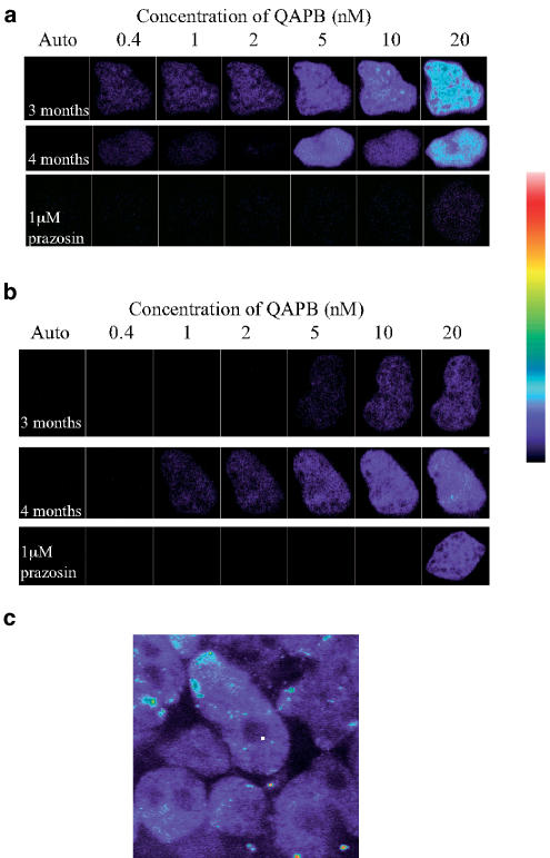
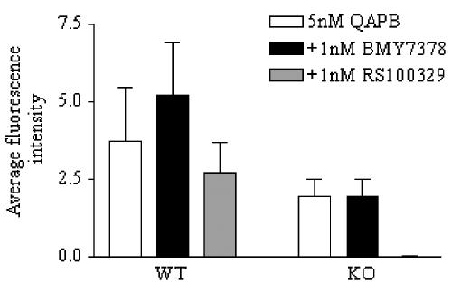
1 Alpha1-adrenoceptors (ARs) play an important functional role in the liver; yet little is known about their cellular location. We identified the subtypes present in wild-type (WT) and alpha1B-AR knockout (KO) mice livers at 3 and 4 months of age, and investigated their distribution in hepatocytes. 2 The fluorescent alpha1-AR antagonist quinazolinyl piperazine borate-dipyrromethene (QAPB) was used to visualise hepatic alpha1-ARs and radioligand binding with [3H]-prazosin was used to quantify the alpha1-AR population. 3 QAPB and [3H]-prazosin bound specifically to hepatic alpha1-ARs with nanomolar affinity. The cellular distribution of alpha1-ARs was similar in WT and alpha1B-AR KO hepatocytes; QAPB binding was distributed diffusely throughout the cell with no binding evident on the plasma membrane. Radioligand binding produced Bmax values as follows: 3-month WT - 76+/-3.3 fmol mg(-1); 4-month WT - 50+/-3.1 fmol mg(-1); 3-month alpha1B-AR KO - 7.4+/-0.73 fmol mg(-1); 4-month alpha1B-AR KO - 30+/-2.0 fmol mg(-1). 4 In 3- and 4-month WT liver, all antagonists acted competitively. RS100329 (alpha1A-selective) and BMY7378 (alpha1D-selective) bound with low affinities, indicating the presence of alpha1B-ARs. In 4-month alpha1B-AR KO liver prazosin produced a biphasic curve, whereas RS100329 and BMY7378 produced monophasic curves of high and low affinity, respectively, indicating the presence of alpha1A-ARs. 5 In conclusion, we have made the novel observation that alpha1-ARs can compensate for one another in the absence of the endogenously expressed receptor; yet there appears to be no subtype-specific subcellular location of alpha1-ARs; the WT livers express alpha1B-ARs, while alpha1B-AR KO livers express alpha1A-ARs. This study provides new insights into both hepatocyte and alpha1-AR biology.
Figures

Comment in
-
Alpha1-adrenoceptor subtype substitution in knockout mice.Br J Pharmacol. 2004 Jul;142(6):919. doi: 10.1038/sj.bjp.0705871. Epub 2004 Jun 21. Br J Pharmacol. 2004. PMID: 15210582 Free PMC article. No abstract available.
References
-
- BYLUND D.B., EIKENBERG D.C., HIEBLE J.P., LANGER S.Z., LEFKOWITZ R.J., MINNEMAN K.P., MOLLINOF P.B., RUFFOLO R.R., JR, TRENDELENBURG U. International Union of Pharmacology: nomenclature of ARs. Pharmacol. Rev. 1994;46:121–136. - PubMed
-
- CAVALLI A., LATTION A.-L., HUMMLER E., NENNIGER M., PEDRAZZINI T., AUBERT J.-F., MICHEL M.C., YANG M., LEMBO G., VECCHIONE C., MOSTARDININ M., SCMIDT A., BEERMANN F., COTECCHIA S. Decreased blood pressure response in mice deficient of the α1b-AR. Proc. Natl. Acad. Sci. U.S.A. 1997;94:11589–11594. - PMC - PubMed
-
- CHALATHORN D., MCCUNE D.F., EDELMANN S.E., GARCIA-CAZARIN L., TSUJIMOTO G., PIASCIK M.T. Differences in the cellular localisation and agonist-mediated internalisation properties of the α1-AR subtypes. Mol. Pharmacol. 2002;61:1008–1116. - PubMed
-
- DALY C.J., DEIGHAN C., MCGEE A., MENNIE D., ALI Z., MCBRIDE M., MCGRATH J.C. A knockout approach indicates a minor vasoconstrictor role for vascular α1B-ARs in mouse. Physiol. Genomics. 2002;9:85–91. - PubMed
-
- DALY C.J., MILLIGAN C.M., MILLIGAN G., MACKENZIE J.F., MCGRATH J.C. Cellular localization and pharmacological characterization of functioning α1-ARs by fluorescent ligand binding and image analysis reveals identical binding properties of clustered and diffuse populations of receptors. J. Pharmacol. Exp. Ther. 1998;286:984–990. - PubMed
Publication types
MeSH terms
Substances
LinkOut - more resources
Full Text Sources
Molecular Biology Databases
Research Materials
