. 2004 Aug 27;279(35):36855-64.
doi: 10.1074/jbc.M405367200. Epub 2004 Jun 21.

Nucleotide release provides a mechanism for airway surface liquid homeostasis

Affiliations

Nucleotide release provides a mechanism for airway surface liquid homeostasis

Eduardo R Lazarowski et al. J Biol Chem. .

Abstract

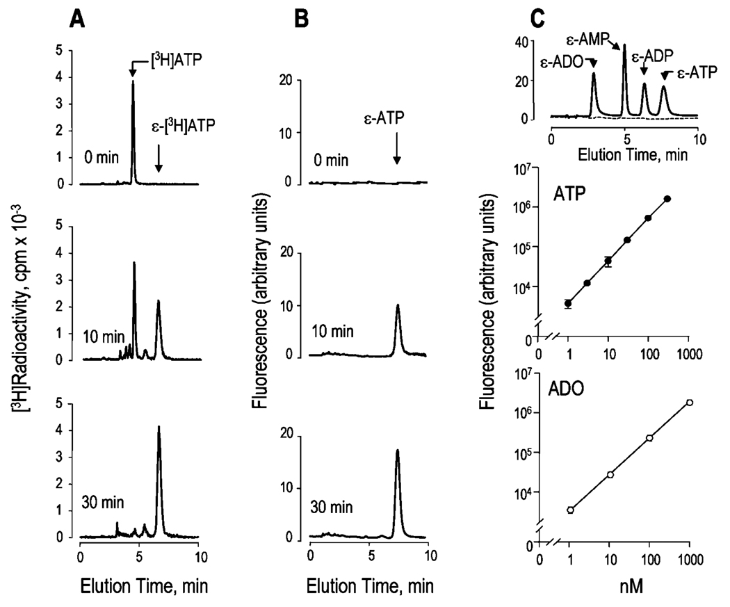
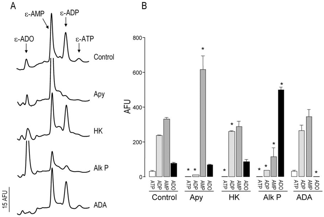
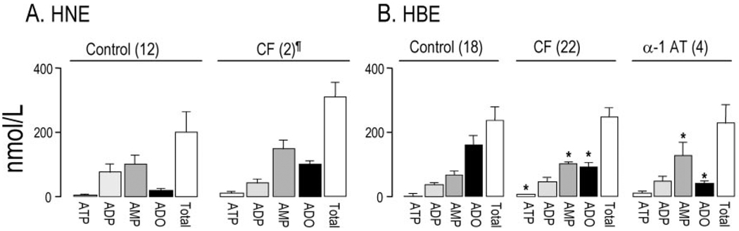
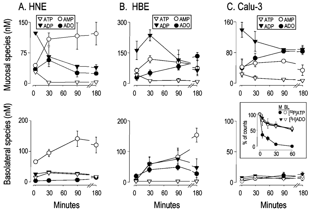
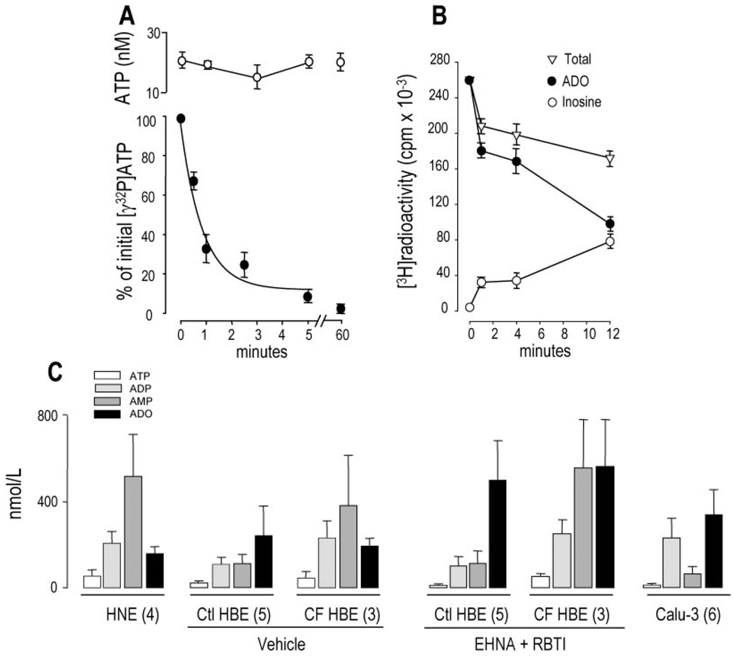
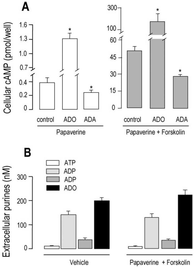
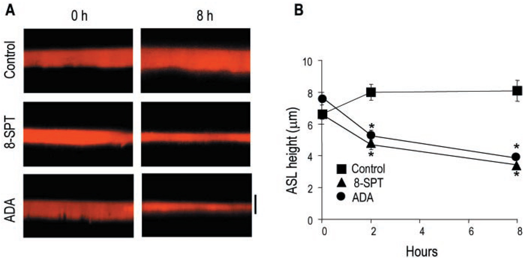
Nucleotides within the airway surface liquid (ASL) regulate airway epithelial ion transport rates by Ca(2+) -and protein kinase C-dependent mechanisms via activation of specific P2Y receptors. Extracellular adenine nucleotides also serve as precursors for adenosine, which promotes cyclic AMP-mediated activation of the cystic fibrosis transmembrane regulator chloride channel via A(2b) adenosine receptors. A biological role for extracellular ATP in ASL volume homeostasis has been suggested by the demonstration of regulated ATP release from airway epithelia. However, nucleotide hydrolysis at the airway surface makes it difficult to assess the magnitude of ATP release and the relative abundance of adenyl purines and, hence, to define their biological functions. We have combined ASL microsampling and high performance liquid chromatography analysis of fluorescent 1,N(6)-ethenoadenine derivatives to measure adenyl purines in ASL. We found that adenosine, AMP, and ADP accumulated in high concentrations relative to ATP within the ASL covering polarized primary human normal or cystic fibrosis airway epithelial cells. By using immortalized epithelial cell monolndogenayers that eously express a luminal A(2b) adenosine receptor, we found that basal as well asforskolin-promoted cyclic AMP production was reduced by exogenous adenosine deaminase, suggesting that A(2b) receptors sense endogenous adenosine within the ASL. The physiological role of adenosine was further established by illustrating that adenosine removal or inhibition of adenosine receptors in primary cultures impaired ASL volume regulation. Our data reveal a complex pattern of nucleotides/nucleosides in ASL under resting conditions and suggest that adenosine may play a key role in regulating ASL volume homeostasis.


Figures

Fig. 1
Fig. 1. Derivatization of adenyl purines
A, conversion of 100 nm [3H]ATP to ε-[3H]ATP was monitored by liquid scintillation detector-coupled HPLC. B, fluorescent HPLC tracings corresponding to the derivatization reaction shown in A. C, fluorescent HPLC tracings from a derivatization reaction performed either in the absence (dashed line) or in the presence (solid line) of 100 nm ADO, AMP, ADP, and ATP (top), and calibration curves for ATP (middle), and adenosine (bottom). All reactions were at 72 °C in the presence of 1 m chloroacetaldehyde, and they took place for the times indicated (A and B) or for 30 min (C). The elution times of authentic [3H]ATP and ε-purine standards are indicated with arrows. HPLC tracings are representative of three (A and B) or at least 20 (C) independent experiments. The data in the middle and bottom panels of C indicate the mean value (±S.D.) from one experiment with triplicate samples, and the results were representative of at least five impendent calibration curves performed under similar conditions.
Fig. 2
Fig. 2. Enzymatic identification of ε-species in ASL
Samples collected from 300-µl mucosal baths of primary cultures of normal resting HNE cells were preincubated with vehicle or with 2 units/ml of the indicated enzyme for 15 min at 30 °C prior to derivatization. A, HPLC tracings. B, changes in fluorescent species (mean ± S.D., n = 3). * indicates significantly different from control. Apy, apyrase; HK, hexokinase, Alk P, alkaline phosphatase; ADA, adenosine deaminase; AFU, arbitrary fluorescence units.
Fig. 3
Fig. 3. Extracellular accumulation of adenyl purines on airway epithelial cells
Confluent cultures of HNE (A) and HBE (B) cells were grown in air/liquid interface and incubated for 3 h with 300 µl of HBSS added bilaterally. The bulk content of the adenyl species was determined by etheno derivatization, as indicated under “Experimental Procedures.” The data represent the mean value ± S.E., or ¶ indicates the mean value ± difference between duplicates; sample number is indicated in parentheses. * indicates significantly different from control cells.
Fig. 4
Fig. 4. Bilateral accumulation of adenyl purines
Well differentiated HNE (A) and HBE cells (B), and Calu-3 cell monolayers (C) were rinsed and incubated in 0.3 ml of bilateral HBSS, and 200 µl were collected at 2, 30, 90, or 180 min and derivatized. Trace amounts (0.1 µCi) of [γ-32P]ATP or [3H]adenosine were added for the indicated times to 0.3 ml of HBSS bathing the mucosal (M) and basolateral (BL) surfaces of Calu-3 cells (C, inset). Fluorescent and radioactive species were analyzed by HPLC, as indicated under “Experimental Procedures.” The data indicate the mean value (±S.D., n = 3) from a single experiment that was representative of two independent experiments performed under similar conditions. Mass measurements at t = 0 (i.e. nominally purine-free HBSS) are not depicted.
Fig. 5
Fig. 5. Basal accumulation of adenyl purines in small volume airway surface liquid
The mucosal surfaces of well differentiated primary HNE and HBE cells and Calu-3 cells were rinsed and preincubated for either 1 (A and B) or 3 h (C) with 25 µl of HBSS. For radiotracer measurements (A and B), 0.1–0.5 µCi of the radiolabeled species were added to the lumen of HBE cultures, and ASL radioactivity was recovered at the times indicated by rapidly rinsing the mucosal cell surface with an excess volume of ice-cold HBSS containing 100 µm of the corresponding nonradioactive purine. [γ-32P]ATP (A, filled circles) and [3H]adenosine (B) were quantified by HPLC as described under “Experimental Procedures.” For ATP measurements by the luciferin-luciferase assay (A, open circles), and for derivatization reactions (C), a micromanipulator was used to collect 5–10 µl of ASL, as described under “Experimental Procedures.” The data in A and B represent the mean value (± S.D.) from at least four independent determinations. C, up to five samples from parallel incubations were pooled and derivatized, and the resulting etheno-adenyl purines were quantified by HPLC. The results are expressed as mean ± S.E.; the number of derivatization reactions is indicated in parentheses. Vehicle or 50 µm erythro-9-(2-hydroxy-3-nonyl)-adenine (EHNA) and 1 µm S-(p-nitrobenzyl)-6-thioinosine (RBTI) were added into the 25 µl of ASL at the beginning of the 3-h incubation period. Ctl, control.
Fig. 6
Fig. 6. Cyclic AMP accumulation and nucleotide release in Calu-3 cells
A, polarized cells (0.9 million cells/Transwell) were rinsed and preincubated for 90 min with 300 µl of mucosal HBSS and 500 µl of basolateral Dulbecco’s modified Eagle’s medium. A, 200 µm papaverine was added in the absence or presence of 5 units/ml adenosine deaminase (ADA). After 10 min, 100 µm ADO and either vehicle (empty bars, left) or 30 µm forskolin (filled bars, right) was added for an additional 10 min as indicated. B, the cells were either incubated for 20 min with vehicle or they were preincubated for 10 min with 200 µm papaverine followed by the addition of 30 µm forskolin and preincubation for 10 min. Extracellular purine and cellular cyclic AMP measurements were performed as described under “Experimental Procedures.” With the exception of papaverine, which was added bilaterally, all other additions were to the mucosal bath. The data indicate the mean value (±S.D.) from triplicate samples, and the results are representative of two experiments performed in identical conditions. *, significantly different from control.
Fig. 7
Fig. 7. Regulation ASL height by endogenous adenosine
A, X-Z confocal images of ASL at 0 and 8 h after mucosal addition of Texas Red dextran in the absence (control) or in the presence of 100 µm 8-SPT or 5 units/ml adenosine deaminase (ADA) to primary HBE cell cultures. The bar indicates 7 µm. B, the data represent the mean ± S.E. (n = 5). *, significantly different from control.

References

    1. Knowles MR, Boucher RC. J. Clin. Investig. 2002;109:571–577. - PMC - PubMed
    1. Boucher RC. Adv. Drug Delivery Rev. 2002;54:1359–1371. - PubMed
    1. Gabriel SE. In: Current Topics in Membranes: Calcium-activated Chloride Channels. Fuller CM, editor. San Diego: Academic Press; 2002. pp. 193–207.
    1. Donaldson SH, Lazarowski ER, Picher M, Knowles MR, Stutts MJ, Boucher RC. Mol. Med. 2000;6:969–982. - PMC - PubMed
    1. Lazarowski ER, Boucher RC, Harden TK. J. Biol. Chem. 2000;275:31061–31068. - PubMed

Publication types

MeSH terms