. 2004 Jun 21;165(6):857-67.
doi: 10.1083/jcb.200401133.

The LIM domain protein UNC-95 is required for the assembly of muscle attachment structures and is regulated by the RING finger protein RNF-5 in C. elegans

Affiliations

The LIM domain protein UNC-95 is required for the assembly of muscle attachment structures and is regulated by the RING finger protein RNF-5 in C. elegans

Limor Broday et al. J Cell Biol. .

Abstract

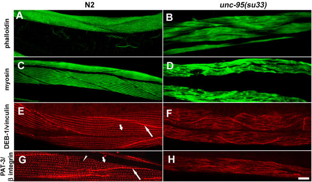
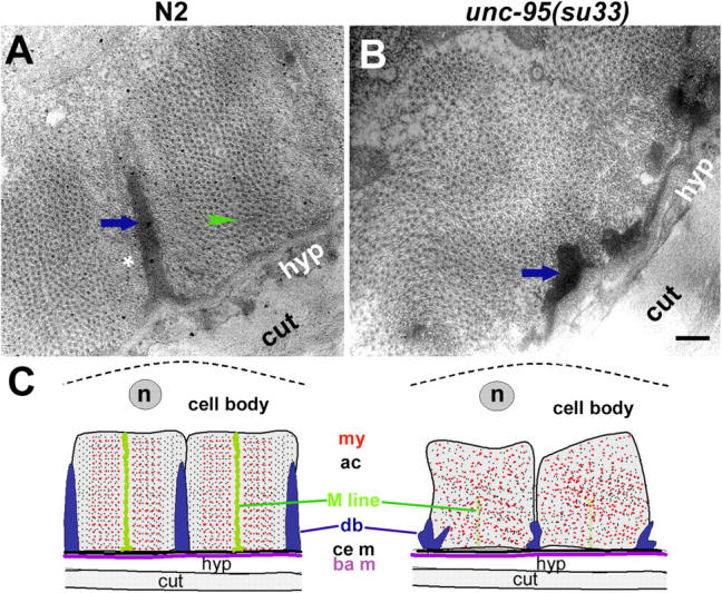
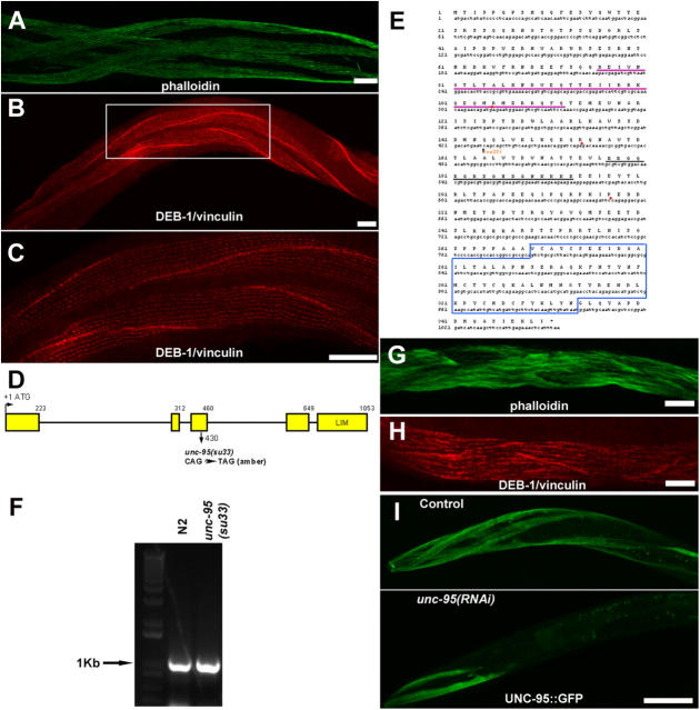
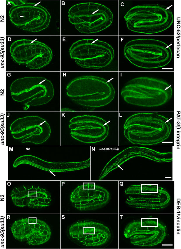
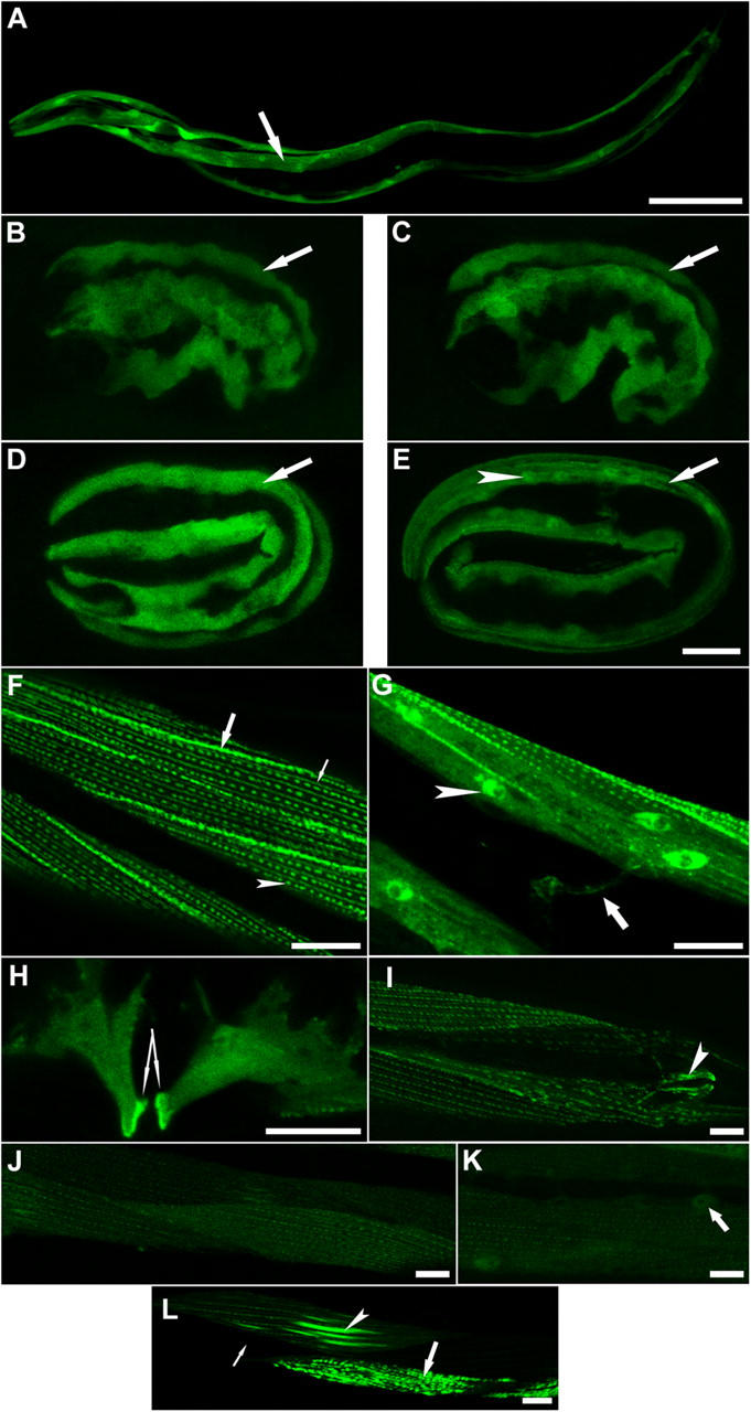
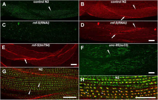
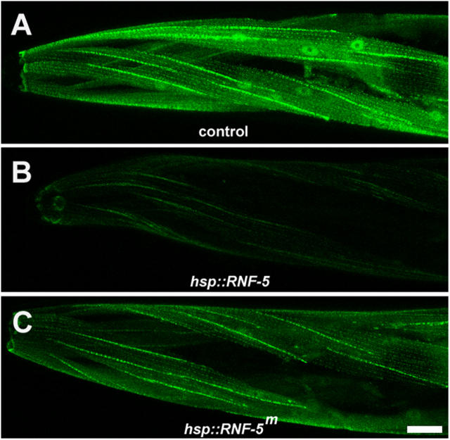
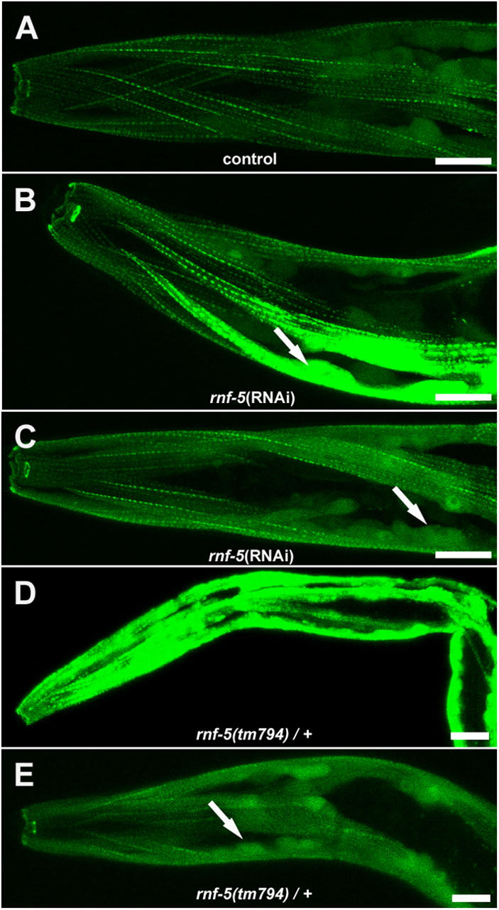
Here, we describe a new muscle LIM domain protein, UNC-95, and identify it as a novel target for the RING finger protein RNF-5 in the Caenorhabditis elegans body wall muscle. unc-95(su33) animals have disorganized muscle actin and myosin-containing filaments as a result of a failure to assemble normal muscle adhesion structures. UNC-95 is active downstream of PAT-3/beta-integrin in the assembly pathways of the muscle dense body and M-line attachments, and upstream of DEB-1/vinculin in the dense body assembly pathway. The translational UNC-95::GFP fusion construct is expressed in dense bodies, M-lines, and muscle-muscle cell boundaries as well as in muscle cell bodies. UNC-95 is partially colocalized with RNF-5 in muscle dense bodies and its expression and localization are regulated by RNF-5. rnf-5(RNAi) or a RING domain deleted mutant, rnf-5(tm794), exhibit structural defects of the muscle attachment sites. Together, our data demonstrate that UNC-95 constitutes an essential component of muscle adhesion sites that is regulated by RNF-5.


Figures

Figure 1.
Figure 1.
Analysis of muscle structure in unc-95(su33) mutant adult animals. Left column shows wild-type staining; right column shows unc-95(su33) staining. (A and B) Actin filament organization in body wall muscles visualized by staining with phalloidin. (C and D) Anti-myosin staining of body wall muscles. (E and F) Anti-DEB-1/vinculin staining of muscle dense bodies and muscle–muscle cell boundaries. Short arrow indicates the muscle dense bodies. Long arrow indicates the muscle–muscle cell boundaries. (G and H) Anti PAT-3/β-integrin staining of dense bodies, M-lines, and muscle–muscle cell boundaries. Short and long arrows are as in E. Arrowhead indicates the muscle M-lines. It is not possible to distinguish between the dense bodies and M-line structures in the unc-95(su33) mutant. Bar, 10 μm.
Figure 2.
Figure 2.
EM of unc-95(su33) adult animals. EM of body wall muscles from wild-type (A) and unc-95(su33) mutant (B). Shown are cross sections of the myofilament lattice; thin and thick filaments appear as dots. Arrows indicate the dense body, arrowheads indicate the M-line, and the asterisk indicates the I band region of thin filaments. Bar, 200 nm. (C) Schematic diagram of cross section of the body wall muscle of wild type and the unc-95(su33) mutant based on the EM data. The body wall muscle cells are associated through the basement membrane (ba m) to the hypodermis (hyp) and cuticle (cut). Dense bodies (db) and M-lines project from the cell membrane (ce m) into the cell and attach actin-containing thin filaments (ac) and myosin-containing thick filaments (my), respectively, to the cell membrane (ce m). The muscle cell body is shown above the sarcomeres including the nucleus (n). The disrupted dense bodies and M-lines of the mutant cause the disorganization of the sarcomeres in the unc-95(su33) mutant.
Figure 3.
Figure 3.
unc-95 encodes a LIM domain protein. (A and B) Rescue of unc-95(su33) mutant animals with the UNC-95::GFP translation fusion construct. Phalloidin staining (A), anti DEB-1/vinculin staining (B), and enlargement of inset in B (C). Use of the rol reporter in the rescue experiments enables the identification of the rescued animals. Bars, 10 μm. (D) Structure of the unc-95 gene (Y105E8A.6). Exons are in yellow. (E) The sequence of unc-95 cDNA and protein. Note the mutation in the su33 allele, the LIM domain (marked in blue) at the COOH terminus, the coiled-coil domain (underlined in purple), and the putative NLS (underlined in black). Introns are labeled in red dots. The sequence data are available from GenBank/EMBL/DDBJ under accession no. NM_170938. Comparison of the LIM domain of UNC-95 to other LIM domain proteins in C. elegans revealed 35% homology to the fourth LIM domain of C28H8.6 (additional C. elegans homologue of vertebrate paxillin, which includes four LIM domains, but not the NH2-terminal extension characteristic of the paxillin family members), 30% homology to the UNC-97 LIM domains, and 30% homology to the predicted protein MLP-1 (T04C9.4b), which includes only one LIM domain and belongs to the MLP/CRP family of muscle LIM domain proteins. (F) RT-PCR analysis of mixed stage RNA purified from N2 and unc-95(su33) cultures. The full-length unc-95 cDNA is 1053 bp. (G and H) unc-95(RNAi) analysis performed on wild-type animals. (G) Phalloidin staining. (H) Anti DEB-1/vinculin staining. Note the phenotype similarity to the su33 allele in Fig. 1 (B and F). Bars, 10 μm. (I) unc-95(RNAi) analysis performed on Ex[unc-95::GFP; rol-6] transgenic animals. Residual GFP expression in body wall muscle cells in the anterior region is shown. In >50% of the RNAi animals analyzed (n = 100), no GFP expression was detected. In all images anterior is to the left. Bars, 50 μm.
Figure 4.
Figure 4.
Muscle assembly during embryogenesis of unc-95(su33) animals. (A–F) Anti-UNC-52/perlecan and MH27 (arrowhead indicates hypodermal cells junctions) staining of wild-type (A–C) and unc-95(su33) (D–F) in 1.5-, 2-, and 3-fold embryos (left, middle, and right panels, respectively). (G–L) Anti-PAT-3/β-integrin staining of wild type (G–I) and unc-95(su33) (J–L). Arrows indicate the pattern of staining in one body wall muscle quadrant. The images show a lateral or dorso-lateral view of the embryo. (M) Wild-type and unc-95(su33) (N) L1 larvae stained with anti-PAT-3/β-integrin. Note the random distribution of dots and lines associated with the muscle dense bodies and M-lines at the L1 stage. (O–T) Anti-DEB-1/vinculin and MH27 staining of wild-type (O–Q) and unc-95(su33) embryos (R–T). Note the disorganized staining and the cytoplasmic accumulation of vinculin in the mutant embryos. Insets are used to indicate the pattern of staining in one muscle quadrant and to highlight the difference in organization between the wild-type and unc-95(su33) mutant embryo. All panels show lateral view of the embryo. Bars, 10 μm.
Figure 5.
Figure 5.
UNC-95 is expressed in muscle attachment sites. (A) Expression of the transcriptional fusion reporter including 2.5 Kb of unc-95 upstream sequences. Arrow points at one of the four body wall muscle quadrants. Bar, 50 μm (B–E). Expression of the translational fusion UNC-95::GFP in developing embryos. The cytoplasmic expression is maintained through embryogenesis. Localization to the muscle attachment sites is shown in E. Arrow indicates one muscle quadrant. Arrowhead indicates the nucleus. Lateral view of the embryo is shown. Bars, 10 μm. (F and G) Expression of the translational fusion UNC-95::GFP in the body wall muscles of adults. (F) UNC-95 is expressed in muscle dense bodies (arrowhead), in M-lines (thin arrow), and in muscle–muscle cell boundaries (thick arrow). (G) UNC-95 localization in the nucleus and cytoplasm of body wall muscles. Arrowhead indicates the nucleus. Expression is also detected in muscle arms (arrow). (H) UNC-95 is localized in the vulval muscles and is highly concentrated in the sites of muscle attachments to the hypodermis (two arrows indicate the strong GFP labeling in the region of attachment). (I) Localization to the anal depressor muscle (arrowhead). (J–L) Expression of the mutant protein unc-95(su33)::GFP translational fusion. (J) Weak staining could be detected in dense bodies and M-lines, but there is no staining in muscle–muscle cell boundaries. (K) Localization of the unc-95(su33)::GFP is maintained in nuclei (arrow). (L) Irregular pattern of staining of the unc-95(su33)::GFP translational fusion. Thin arrow indicates cell in which the transgene was silenced; arrowhead indicates cell in which the M-lines are much thicker; thick arrow indicates cell in which the dense bodies aggregate to abnormal structures. Bars, 10 μm.
Figure 6.
Figure 6.
RNF-5 has a role in the maintenance of muscle dense bodies. (A–D) rnf-5(RNAi) analysis. (A and B) Control animals double stained with the anti-RNF-5 (A; green) and anti-DEB-1/vinculin (B; red) antibodies. (C and D) rnf-5(RNAi) animals. Note weak staining with the RNF-5 antibody in C, demonstrating that the RNAi treatment indeed depleted the endogenous RNF-5. Note the aberrant structure of dense bodies and muscle–muscle cell boundaries in (D). (E) Staining of the rnf-5(tm794) homozygous animals with anti-DEB-1/vinculin antibody. (F) Staining of unc-95(su33) adult muscle with anti-RNF-5 antibody. Note the aberrant dense body structure as compared with A. (G) RNF-5 (green) and DEB-1/vinculin (red) are localized in close proximity in the muscle dense bodies. (H) Partial colocalization (yellow) of RNF-5 (red) with GFP (green) of the UNC-95::GFP expression construct in muscle dense bodies. Arrowheads indicate the dense bodies. Arrows indicate muscle–muscle cell boundaries. Bars, 10 μm.
Figure 7.
Figure 7.
Ectopic expression of RNF-5 in the UNC-95::GFP transgenic animals affects GFP expression levels. (A) Control level of expression of the translational fusion UNC-95::GFP. (B) Overexpression of wild-type RNF-5 under the heat shock promoter. UNC-95::GFP levels are lower compared with control. (t test; P < 0.005, n = 10; based on measurement of GFP fluorescence). (C) Overexpression of a RING mutant form of RNF-5 (RNF-5m) under the heat shock promoter. Note weaker effect of the mutant form on GFP levels (t test; P < 0.05, n = 10; based on measurement of GFP fluorescence). The representative images shown were captured using the same confocal parameters; all panels were enhanced in order to enable the detection of the expression pattern in B. Bar, 10 μm.
Figure 8.
Figure 8.
rnf-5(RNAi) and rnf-5(tm794) heterozygous caused accumulation of the UNC-95::GFP transgene in muscle cell bodies. (A) Control level of expression of the translational fusion UNC-95::GFP. (B and C) Two representative images of rnf-5(RNAi) animals. The muscle cell body is indicated by an arrow. Note increased GFP expression, especially in muscle cell body. (D and E) Two representative images of rnf-5(tm794) heterozygous animals. Arrow as in C. All images were captured using the same confocal parameters. Bars, 10 μm.

References

    1. Bach, I. 2000. The LIM domain: regulation by association. Mech. Dev. 91:5–17. - PubMed
    1. Barstead, R.J., and R.H. Waterston. 1991. Vinculin is essential for muscle function in the nematode. J. Cell Biol. 114:715–724. - PMC - PubMed
    1. Benian, G.M., T.L. Tinley, X. Tang, and M. Borodovsky. 1996. The Caenorhabditis elegans gene unc-89, required for muscle M-line assembly, encodes a giant modular protein composed of Ig and signal transduction domains. J. Cell Biol. 132:835–848. - PMC - PubMed
    1. Dawid, I.B., J.J. Breen, and R. Toyama. 1998. LIM domains: multiple roles as adapters and functional modifiers in protein interactions. Trends Genet. 14:156–162. - PubMed
    1. Didier, C., L. Broday, A. Bhoumik, S. Israeli, S. Takahashi, K. Nakayama, S.M. Thomas, C.E. Turner, S. Henderson, H. Sabe, and Z. Ronai. 2003. RNF5, a RING finger protein that regulates cell motility by targeting paxillin ubiquitination and altered localization. Mol. Cell. Biol. 23:5331–5345. - PMC - PubMed

Publication types

MeSH terms

Substances