Comparative Study
. 2004 Jun 21;165(6):881-91.
doi: 10.1083/jcb.200403174.

Melanoma chondroitin sulfate proteoglycan enhances FAK and ERK activation by distinct mechanisms

Affiliations
Comparative Study

Melanoma chondroitin sulfate proteoglycan enhances FAK and ERK activation by distinct mechanisms

Jianbo Yang et al. J Cell Biol. .

Abstract

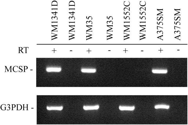
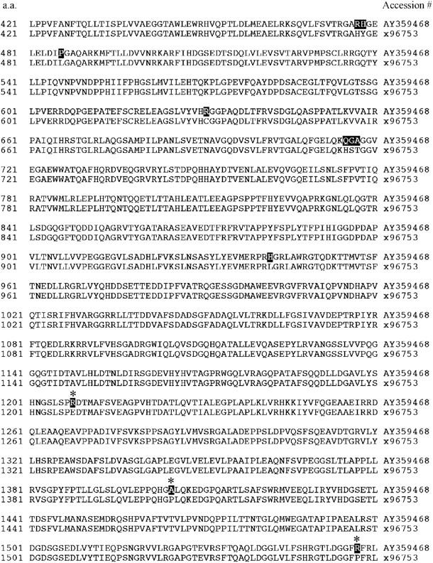
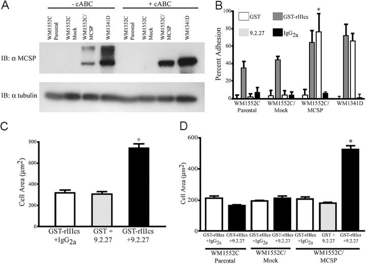
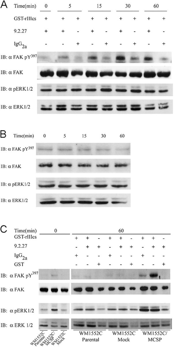
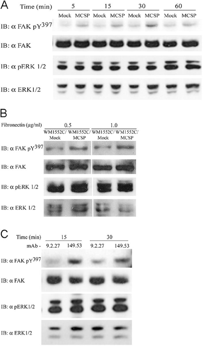
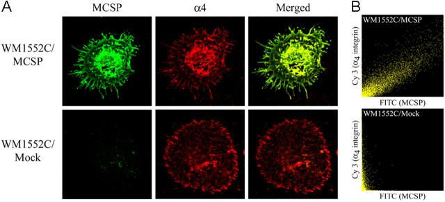
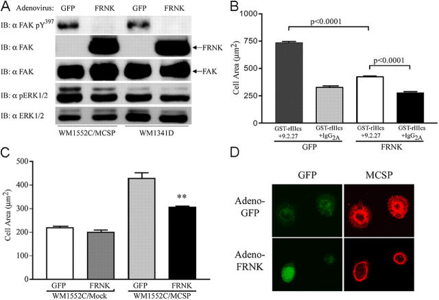
Melanoma chondroitin sulfate proteoglycan (MCSP) is an early cell surface melanoma progression marker implicated in stimulating tumor cell proliferation, migration, and invasion. Focal adhesion kinase (FAK) plays a pivotal role in integrating growth factor and adhesion-related signaling pathways, facilitating cell spreading and migration. Extracellular signal-regulated kinase (ERK) 1 and 2, implicated in tumor growth and survival, has also been linked to clinical melanoma progression. We have cloned the MCSP core protein and expressed it in the MCSP-negative melanoma cell line WM1552C. Expression of MCSP enhances integrin-mediated cell spreading, FAK phosphorylation, and activation of ERK1/2. MCSP transfectants exhibit extensive MCSP-rich microspikes on adherent cells, where it also colocalizes with alpha4 integrin. Enhanced activation of FAK and ERK1/2 by MCSP appears to involve independent mechanisms because inhibition of FAK activation had no effect on ERK1/2 phosphorylation. These results indicate that MCSP may facilitate primary melanoma progression by enhancing the activation of key signaling pathways important for tumor invasion and growth.


Figures

Figure 1.
Figure 1.
Expression of MCSP mRNA in melanoma cell lines by RT-PCR. Poly(A)+ RNA was isolated from each of the cell lines indicated and equal amounts were reverse transcribed (± reverse transcriptase), followed by PCR amplification with primers specific for MCSP or glyceraldehyde-3-phosphate dehydrogenase (G3PDH), as a control.
Figure 2.
Figure 2.
Partial amino acid sequence alignment of the MCSP cDNA generated from A375SM melanoma cells and the published sequence. Highlighted sequences represent revisions of the original sequence (Pluschke et al., 1996). The new and original sequence data are available from GenBank/EMBL/DDBJ under accession no. AY359468 and X96753, respectively. Asterisks represent novel amino acid changes not previously reported for MCSP or NG2.
Figure 3.
Figure 3.
MCSP supports adhesion and induces spreading of WM1552C/MCSP-transfected cells. (A) WM1552C cells were stably transfected with MCSP (WM1552C/MCSP) and expression verified by immunoblot. Whole-cell lysates from cells incubated ±0.5 U/ml cABC were fractionated by SDS-PAGE and probed with anti-MCSP core protein mAb 9.2.27. (B) Cells were plated in 96-well plates on 3 μg/ml GST, 3 μg/ml GST-rIIIcs, 1 μg/ml mAb 9.2.27, or 1 μg/ml IgG2a, allowed to adhere for 30 min at 37°C, and the number of adherent cells was determined by formazan absorbance. Data shown represent the mean of triplicate wells, ± SD. (C) WM1341D- and (D) WM1552C-transfected cells were serum starved overnight and plated on chimeric substrata using the same concentrations as in B. Plates were incubated at 37°C for 1 h, washed, fixed, and stained as described in the Materials and methods. Cell areas of a random 50 cells/well from triplicate wells were quantified by tracing the cell border using NIH Image software. *, P < 0.001 by two-tailed t test.
Figure 4.
Figure 4.
MCSP stimulates FAK Y 397 and ERK1/2 phosphorylation in melanoma cells. Cells were serum starved overnight and plated on various chimeric substrata using the same concentrations described in Fig. 3 B. (A) WM1341D cells were allowed to adhere to the specified substrata at 37°C for the indicated times, lysed with SDS sample buffer, and immunoblotted with total and phosphospecific FAK and ERK1/2 antibodies as indicated. (B) WM1341D cells were allowed to adhere to plates coated with mAb 9.2.27 and GST for the indicated times at 37°C, lysed in SDS sample buffer, and lysates were evaluated by immunoblotting as in A. (C) WM1552C parental and transfectant cells were plated on the indicated substrata and allowed to adhere for 1 h at 37°C. Cells were lysed in SDS sample buffer and analyzed for levels of FAK and ERK1/2 phosphorylation as in A.
Figure 5.
Figure 5.
Plating on GST-FN51 or FN promotes FAK Y 39 7 phosphorylation. WM1552C/Mock and WM1552C/MCSP cells were plated at 37°C in 35-mm Petri dishes coated with 0.5 μg/ml GST-FN51 for the indicated times (A), or with FN for 1 h (B) in SIFM. The cells were lysed by addition of SDS sample buffer and the lysates were evaluated for FAK and ERK1/2 phosphorylation/expression by immunoblotting as indicated. White lines indicate that intervening lanes have been spliced out. (C) Serum-starved WM1552C/MCSP cells were released and pretreated with 5 μg/ml of either anti-MCSP mAb 9.2.27 or mAb 149.53 for 30 min at 37°C. Cells were plated on FN51-coated dishes (0.5 μg/ml) for the indicated times at 37°C, lysed in SDS sample buffer, and analyzed by immunoblotting with the indicated antibodies.
Figure 6.
Figure 6.
MCSP and α4 integrin colocalize upon engagement on GST-FN51. (A) WM1552C/Mock and MCSP cells were plated on coverslips coated with 5 μg/ml GST-FN51 for 1 h at 37°C in SIFM, and were then fixed. Cells were double stained with polyclonal anti-MCSP antibody (FITC) and anti-α4 integrin mAb (Cy3), followed by the appropriate secondary antibody as indicated. The colocalization of these receptors upon engagement is evident in the merged images (yellow). (B) Z sections from the adherent boundary of the cell were analyzed for the colocalization of MCSP (FITC) and α4 integrin (Cy3) using the Fluoview™ software colocalization processor. Pixel pairs demonstrating staining for both receptors plot at a 45° angle on the scattergram (WM1552C/MCSP cells), whereas pixels staining with only one fluorophore fall along the corresponding axis (WM1552C/Mock cells).
Figure 7.
Figure 7.
Overexpression of dominant-negative FAK (FRNK) inhibits FAK phosphorylation and MCSP-mediated melanoma cell spreading. (A) Cells were infected in SIFM with an adenoviral FRNK/GFP construct or an adenoviral construct expressing only GFP at 37°C overnight, released, and allowed to adhere on plates coated with GST-rIIIcs/mAb 9.2.27 for 1 h at 37°C. Cell lysates were analyzed for tyrosine phosphorylation of FAK Y397 by immunoblotting. (B) WM1341D melanoma cells were infected as in A, allowed to spread for 1 h at 37°C in wells coated with GST-rIIIcs/mAb 9.2.27 or GST-rIIIcs/IgG2a, and the cell areas were measured as described in the Materials and methods. (C) WM1552C/Mock and MCSP-transfected cells were infected overnight as in A. Cells were plated in wells coated with GST-rIIIcs/9.2.27 for 1 h at 37°C, and the adherent cells were measured as described in the Materials and methods. **, P < 0.001 by two-tailed t test. (D) WM1552C/MCSP cells infected with either adeno-GFP or adeno-FRNK were plated on coverslips coated with 5 μg/ml GST-FN51, fixed, and stained for MCSP. Representative images from confocal analysis are shown for GFP (green) and MCSP staining (red).

References

    1. Aplin, A.E., and R.L. Juliano. 1999. Integrin and cytoskeletal regulation of growth factor signaling to the MAP kinase pathway. J. Cell Sci. 112:695–706. - PubMed
    1. Barberis, L., K.K. Wary, G. Fiucci, F. Liu, E. Hirsch, M. Brancaccio, F. Altruda, G. Tarone, and F.G. Giancotti. 2000. Distinct roles of the adaptor protein Shc and focal adhesion kinase in integrin signaling to ERK. J. Biol. Chem. 275:36532–36540. - PubMed
    1. Bogenrieder, T., and M. Herlyn. 2002. Cell-surface proteolysis, growth factor activation and intercellular communication in the progression of melanoma. Crit. Rev. Oncol. Hematol. 44:1–15. - PMC - PubMed
    1. Burg, M.A., K.A. Grako, and W.B. Stallcup. 1998. Expression of the NG2 proteoglycan enhances the growth and metastatic properties of melanoma cells. J. Cell. Physiol. 177:299–312. - PubMed
    1. Burridge, K., and M. Chrzanowska-Wodnicka. 1996. Focal adhesions, contractility, and signaling. Annu. Rev. Cell Dev. Biol. 12:463–518. - PubMed

Publication types

MeSH terms

Substances