. 2004 Jun 21;199(12):1689-700.
doi: 10.1084/jem.20032202.

Early B cell factor promotes B lymphopoiesis with reduced interleukin 7 responsiveness in the absence of E2A

Affiliations

Early B cell factor promotes B lymphopoiesis with reduced interleukin 7 responsiveness in the absence of E2A

Christopher S Seet et al. J Exp Med. .

Abstract

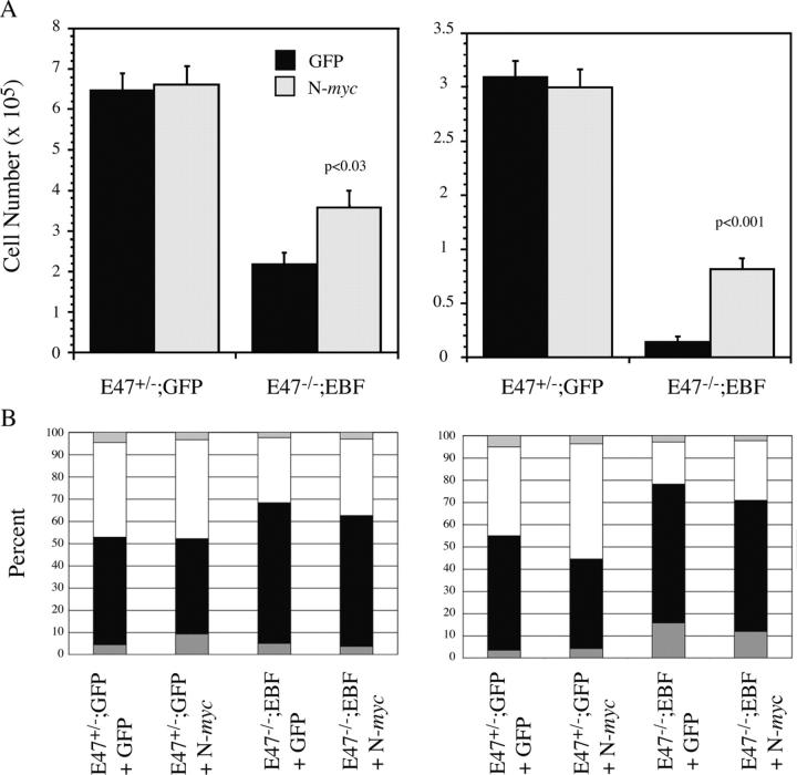
The basic helix-loop-helix transcription factors encoded by the E2A gene function at the apex of a transcriptional hierarchy involving E2A, early B cell factor (EBF), and Pax5, which is essential for B lymphopoiesis. In committed B lineage progenitors, E2A proteins have also been shown to regulate many lineage-associated genes. Herein, we demonstrate that the block in B lymphopoiesis imposed by the absence of E2A can be overcome by expression of EBF, but not Pax5, indicating that EBF is the essential target of E2A required for development of B lineage progenitors. Our data demonstrate that EBF, in synergy with low levels of alternative E2A-related proteins (E proteins), is sufficient to promote expression of most B lineage genes. Remarkably, however, we find that E2A proteins are required for interleukin 7-dependent proliferation due, in part, to a role for E2A in optimal expression of N-myc. Therefore, high levels of E protein activity are essential for the activation of EBF and N-myc, whereas lower levels of E protein activity, in synergy with other B lineage transcription factors, are sufficient for expression of most B lineage genes.


Figures

Figure 1.
Figure 1.
E2A proteins are required for development of BLPs and expression of EBF and Pax5 in FL. (A) FACS® analysis for B220 and CD19 (top) or c-kit (bottom)–expressing cells in the liver E47+/− and E47−/− embryos at E15.5. (B) Semiquantitative RT-PCR analysis of EBF, Pax5, and actin mRNA in Lin cells isolated from E47+/+, E47+/−, or E47−/− FL at E12.5 or E14.5. Threefold serial dilutions of cDNA, starting at 50 ng/reaction were amplified for 32 cycles (EBF), 35 cycles (Pax5), or 28 cycles (actin). In all experiments, RNA that had not been reverse transcribed (−RT) served as a negative control. (C) FACS® analysis of CD19 expression on E47−/−;GFP (dark line), E47+/−;GFP (dotted line), and E47−/−;E2A (light line) FL cells 18 d after infection. (D) RT-PCR analysis of EBF and actin mRNA from E47+/−;GFP, E47−/−;GFP, and E47−/−;E2A cells 15 d after infection. 15 ng cDNA was amplified for 30 cycles (EBF) or 28 cycles (actin). In all RT-PCR experiments, the lanes being compared were amplified in parallel and run on the same gel, although the lanes have been rearranged to aid in comparison.
Figure 2.
Figure 2.
Development of B220+ CD19+ BP1+ cells from E47−/− FL progenitors expressing EBF, but not Pax5. (A) Hematopoietic progenitors were isolated from E47+/− and E47−/− FL cells at day 12 of gestation, infected with the GFP only, EBF, or Pax5-producing retrovirus, and cultured on S17 stromal cells in the presence of KL and IL-7. 12 d after the initiation of culture, the resulting cell populations were isolated and stained with antibodies recognizing CD11b, Gr-1, B220, and CD19. The plots show B220 and CD19 expression on CD11b Gr-1 cells. (B) 18 d after the initiation of culture, the total number of cells that had developed from the starting population was determined. (C) 20 d after the initiation of culture, the indicated cell populations were stained with antibodies recognizing B220, CD19, or BP-1/6C3.
Figure 3.
Figure 3.
Expression of B lineage genes and IgH rearrangements in E47−/−;EBF BLPs. (A) RT-PCR analysis of B lineage gene expression in RNA isolated from E47+/−;GFP, E47−/−;GFP, E47−/−;EBF, and E47−/−;Pax5 populations 20 d after infection. The input cDNA was standardized such that a similar amount of actin was amplified under semiquantitative conditions. In all RT-PCR experiments, the lanes being compared were amplified in parallel and run on the same gel. The +RT and −RT lanes were juxtaposed to aid in comparison. (B) B220+ CD19+ cells were sorted from cultures of E47+/−;GFP and E47−/−;EBF cells and total RNA was extracted. Semiquantitative RT-PCR was performed on serial threefold dilutions of reverse transcribed RNA, 50 ng/reaction, using the indicated PCR primers. In all experiments, RNA that had not been reverse transcribed (−RT) served as a negative control. (C) PCR-amplified products from DNA isolated from B220+ CD19+ E47+/−;GFP (lane 1) and E47−/−;EBF (lane 2) BLPs using primers to detect the germline (GL) configuration of the IgH locus, (D) the JH4 primer in conjunction with the DHL (L) primer, which detects D-JH rearrangements, or the DHR (R) primer, which is in the opposite orientation and should not amplify D-JH rearrangements, and (E) the VJ558, V7183, and VQ52 primers in combination with the JH4 primer. All amplified products were transferred to nylon membrane and probed with a radiolabeled cDNA fragment containing a DJH4 fragment.
Figure 4.
Figure 4.
EBF does not induce E protein mRNA or protein in E47−/−;EBF cells. (A) Total RNA isolated from sorted B220+ CD19+ E47+/−;GFP (open bars) and E47−/−;EBF (solid bars) cells at day 20 of culture was analyzed by real time PCR for expression of E12, E47, HEB, and E2-2. The abundance of target mRNA relative to the level of HPRT mRNA is shown. Error bars represent the standard error of five replicate samples. (B) EMSA of whole cell protein extracts prepared from sorted B220+ CD19+ E47+/−;GFP (lanes 1 and 2) and E47−/−;EBF (lanes 3 and 4) cells 25 d after the initiation of culture. The extracts were incubated with the probe in the presence of a noncompetitive (lanes 1 and 3) or competitive (lanes 2 and 4) oligonucleotide, except in the case of the Oct probe, where no competitive oligonucleotide was added. Lanes 1–4 for each of the probes were run on the same gel. The probes used include the E-box–containing μE5 probe, the EBF site from the mb-1 promoter, a composite Pax5/ets site from the mb-1 promoter, and an Octamer protein binding sequence as indicated at the bottom of the panel.
Figure 5.
Figure 5.
E47−/−;EBF BLPs require E protein activity for survival and expression of RAG-1 and RAG-2. (A) E47+/−;GFP and E47−/−;EBF BLPs were infected with ERId3m (solid bars) or ERId3 (gray bars). 24 h after infection, the cells were treated with 1 μM estradiol for 9 h and the percent of GFPhi cells binding annexin V was determined by flow cytometry. (B) E47+/−;GFP and E47−/−;EBF BLPs were infected with hCD25- or Id3/hCD25-producing retrovirus. 24 h after infection, hCD25+ cells were isolated and total RNA was extracted and reverse transcribed for analysis by semiquantitative RT-PCR. Expression of RAG-1, RAG-2, CD19, and actin are shown. In all RT-PCR experiments, the lanes being compared were amplified in parallel and run on the same gel. The +RT and −RT lanes were juxtaposed after photographing the gel.
Figure 6.
Figure 6.
E47−/−;EBF BLPs proliferate less well than E47+/−;GFP BLPs and have reduced expression of N-myc. (A) 105 B220+ CD19+ cells from E47+/−;GFP (solid bars) and E47−/−;EBF (gray bars) cultures were plated in IL-7 alone or S17 + KL + IL-7. 48 h later, the cells were harvested and counted on the hemacytometer. The standard error of three replicate cultures is indicated. The p-values were generated using a Student's t test. (B) Cell cycle analysis using BrdU and PI of E47+/−;GFP and E47−/−;EBF BLPs cultured on S17 + KL + IL-7. The percentage of cells in each phase of the cell cycle is plotted. The data represent one of two independent experiments. (C) Northern blot analysis of RNA extracted from E47+/−;GFP and E47−/−;EBF BLPs or (D) BLPs expressing ERId3 or ERId3m after treatment with 1 μM 17β-estradiol for 6 h. The probes used are indicated on the right of each panel.
Figure 7.
Figure 7.
N-myc promotes the proliferation of E47−/−;EBF BLPs. (A) E47+/−;GFP and E47−/−;EBF BLPs were infected with GFP (solid bars) or N-myc (gray bars)–producing retrovirus and 48 h later, three replicate wells of 105 cells were plated on S17 + KL + IL-7 (right) or IL-7 alone (left). The total number of cells present in each well was determined 48 h later. The p-values were generated using a Student's t test. (B) 96 h after infection with GFP or N-myc retrovirus, E47+/−;GFP and E47−/−;EBF BLPs were incubated for 20 min with BrdU and then stained with anti-BrdU and PI. The percent of cells in each phase of the cell cycle was determined by flow cytometry.

Similar articles

Cited by

References

    1. Kee, B.L., and C.J. Paige. 1995. Murine B cell development: commitment and progression from multipotential progenitors to mature B lymphocytes. Int. Rev. Cytol. 157:129–179. - PubMed
    1. Kee, B.L., and C. Murre. 2001. Transcription factor regulation of B lineage commitment. Curr. Opin. Immunol. 13:180–185. - PubMed
    1. O'Riordan, M., and R. Grosschedl. 2000. Transcriptional regulation of early B-lymphocyte differentiation. Immunol. Rev. 175:94–103. - PubMed
    1. Bain, G., E.C. Robanus Maandag, D.J. Izon, D. Amsen, A.M. Kruisbeek, B.C. Weintraub, I. Krop, M.S. Schlissel, A.J. Feeney, M. van Roon, et al. 1994. E2A proteins are required for proper B cell development and initiation of immunoglobulin gene rearrangements. Cell. 79:885–892. - PubMed
    1. Zhuang, Y., P. Soriano, and H. Weintraub. 1994. The helix-loop-helix gene E2A is required for B cell formation. Cell. 79:875–884. - PubMed

Publication types

MeSH terms