. 2004 Jun 21;32(11):3304-15.
doi: 10.1093/nar/gkh651. Print 2004.

Efficient downregulation of immunoglobulin mu mRNA with premature translation-termination codons requires the 5'-half of the VDJ exon

Affiliations

Efficient downregulation of immunoglobulin mu mRNA with premature translation-termination codons requires the 5'-half of the VDJ exon

Marc Bühler et al. Nucleic Acids Res. .

Abstract

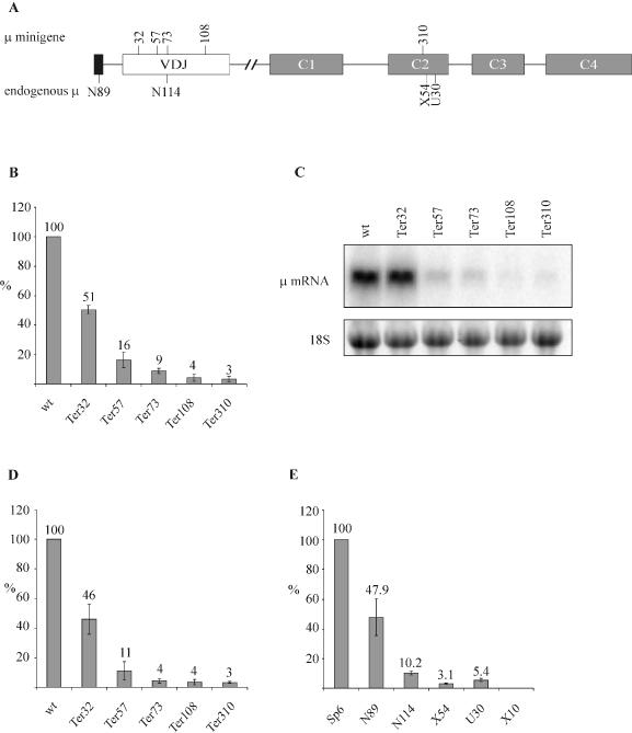
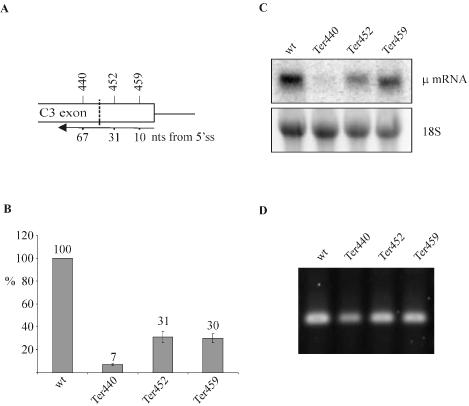
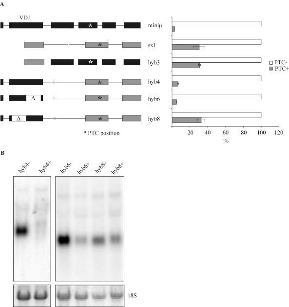
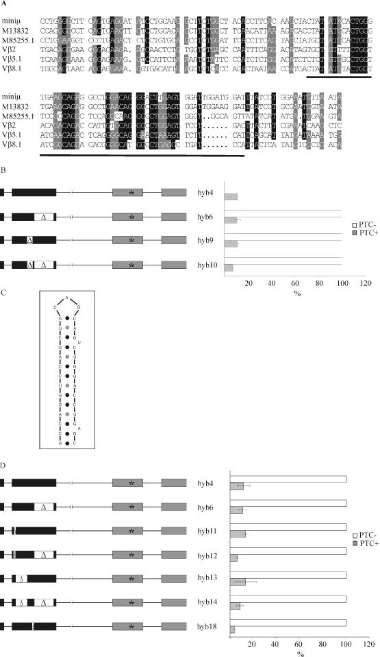
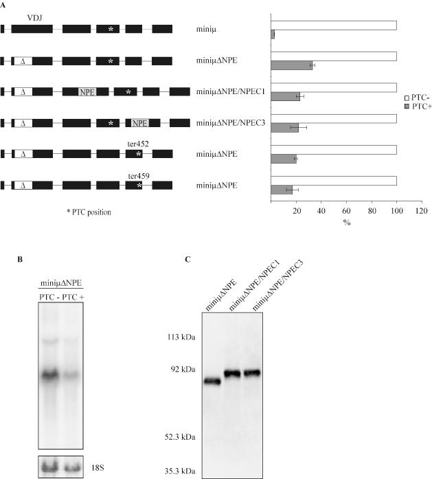
Premature translation-termination codons (PTCs) elicit rapid degradation of the mRNA by a process called nonsense-mediated mRNA decay (NMD). NMD appears to be significantly more efficient for mRNAs of genes belonging to the immunoglobulin superfamily, which frequently acquire PTCs during VDJ rearrangment, than for mRNAs of other genes. To identify determinants for efficient NMD, we developed a minigene system derived from a mouse immunoglobulin micro gene (Ig-micro) and measured the effect of PTCs at different positions on the mRNA level. This revealed that PTCs located downstream of the V-D junction in the VDJ exon of Ig-micro minigenes and of endogenous Ig-micro genes elicit very strong mRNA downregulation, whereas NMD efficiency decreases gradually further upstream in the V segment where a PTC was inserted. Interestingly, two PTCs are in positions where they usually do not trigger NMD (<50 nt from the 3'-most 5' splice site) still resulted in reduced mRNA levels. Using a set of hybrid constructs comprised of Ig-micro and an inefficient substrate for NMD, we identified a 177 nt long element in the V segment that is necessary for efficient downregulation of PTC-containing hybrid transcripts. Moreover, deletion of this NMD-promoting element from the Ig-micro minigene results in loss of strong NMD.


Figures

Figure 1
Figure 1
Efficiency of NMD increases with 5′ to 3′ polarity. (A) Schematic representation of the Ig-μ gene, with numbers above depicting the amino acid positions where PTCs were inserted in the μ-minigene. The position of the PTC in the endogenous Ig-μ gene in the different hybridoma cell lines is shown below the diagram. The original clone names of the hybridoma cell lines are used throughout this paper (22). The PTC in cell line N89, N114, X54 and U30 is located at amino acid positions 3, 73, 325 and 335, respectively. (B) Relative Ig-μ mRNA levels of HeLa cells transiently transfected with the indicated Ig-μ minigenes under control of the human β-actin promotor were analyzed 48 h post transfection by real-time RT–PCR. The indicated relative Ig-μ mRNA levels were normalized to neomycin mRNA encoded on the transfected plasmid. ‘wt’ denotes the PTC-free Ig-μ minigene, the various PTC+ constructs are named ‘Ter’ (for termination) followed by a number indicating the amino acid position of the PTC. (C) Analysis of the same RNA used in (B) by northern blotting using a 32P-labeled probe for Ig-μ mRNA. As a loading control, the 18S rRNA band from the ethidium bromide-stained gel before blotting is shown in the lower panel. (D) HeLa cells were transfected with the same Ig-μ minigene constructs as in (B) and stably transfected polyclonal cell pools were generated by selection with Geneticin. Relative Ig-μ mRNA levels were determined by real-time RT–PCR as in (B) and normalized to relative endogenous GAPDH mRNA levels. (E) RNA of the parental hybridoma cell line Sp6, encoding a productively rearranged full-length Ig-μ mRNA, and of five Sp6-derived, mutated cell clones was analyzed. X10 has deleted the entire Ig-μ gene and serves as a negative control. Relative Ig-μ mRNA levels were determined by real-time RT–PCR as in (B) and normalized to relative 18S ribosomal RNA levels. In (B), (D) and (E), average values of three real-time PCR runs with cDNAs of one representative experiment are shown. Error bars indicate standard deviations.
Figure 2
Figure 2
Testing the ‘50 nucleotides boundary rule’ for NMD of Ig-μ minigenes. (A) PTCs were introduced into exon C3 of the Ig-μ minigene at the amino acid positions indicated above the diagram. The numbers below depict the distance of these PTCs from the 3′-most 5′ splice site. The ‘50 nucleotides boundary’ is marked by the dashed line. (B) RNA of polyclonal HeLa cell pools stably transfected with the indicated Ig-μ minigenes under control of the human β-actin promotor were analyzed by real-time RT–PCR. The indicated relative Ig-μ mRNA levels were normalized to endogenous GAPDH mRNA. Average values of three real-time PCR runs of a typical experiment are shown. Error bars indicate standard deviations. (C) Northern blot analysis of RNA harvested 48 h post transfection with a 32P-labeled probe for Ig-μ mRNA. As a loading control, the 18S rRNA band from the ethidium bromide-stained gel before blotting is shown in the lower panel. (D) RT–PCR analysis of the RNA used in (B) to check for potential cryptic splicing between exon C3 and exon C4. Correct joining of exon C3 to exon C4 is predicted to give a PCR product of 163 bp.
Figure 3
Figure 3
The 5′ half of the Ig-μ minigene VDJ exon is necessary for efficient NMD of Ig-μ/Sxl hybrid transcripts. (A) Relative hybrid mRNA levels of HeLa cells stably expressing the constructs schematically represented on the left were determined by real-time RT–PCR and are shown in the right panel. For each pair of hybrid constructs, the PTC− mRNA level (white bars) was set as 100% and the PTC+ mRNA level (gray bars) was calculated relative to it. The hybrid mRNA values were normalized to endogenous GAPDH mRNA. Average values of three real-time PCR runs of a typical cell pool are shown, and error bars indicate standard deviations. In the left panel, exons originating from Ig-μ are depicted in black and Sxl exons are depicted in gray. The position of the PTC is indicated by an asterisk and corresponds to ter310 in miniμ or ter43 in Sxl, respectively. The white regions labeled with Δ mark deletions. (B) Northern blot analysis of 15 μg of total cellular RNA isolated from HeLa cells stably transfected with the indicated hybrid constructs. As a loading control, the 18S rRNA band from the ethidium bromide-stained gel before blotting is shown in the lower panel.
Figure 4
Figure 4
Smaller deletions within the 5′ half of the Ig-μ VDJ exon did not affect its NMD-promoting activity. (A) Alignment of VDJ exon sequences of several different human and mouse Ig-μ and TCR-β genes. ‘Miniμ’ is from the mouse Ig-μ of the hybridoma cell line Sp6 (23). Accession numbers M13832 and M85255.1 are rearranged VDJ regions of mouse and human Ig-μ, respectively. Vβ2 (accession no. X04331), Vβ5.1 (19) and Vβ8.1 (19) are mouse TCR-β sequences. The underlined sequence corresponding to amino acid positions 52–70 in our Ig-μ minigene contains conserved motifs and is deleted in hyb9 and hyb10. (B) Total cellular RNA of HeLa cells transiently transfected with the indicated Ig-μ/Sxl hybrid genes were analyzed 48 h post transfection by real-time RT–PCR. Relative Ig-μ mRNA levels were normalized to neomycin mRNA encoded on the transfected plasmid. Average values of three real-time PCR runs of a typical experiment are shown. Error bars indicate standard deviations. (C) Predicted stem–loop structure located between amino acid positions 19 and 30 of Ig-μ. (D) The 33 bp comprising the predicted stem–loop structure depicted in (C) were deleted from hyb4 and hyb6, giving hyb11 and hyb12, respectively. The 63 bp between the predicted stem–loop and the underlined motif in (A) were deleted from hyb4 and hyb6, giving hyb13 and hyb14, respectively. The 18 bp comprising amino acid positions 71–76 were deleted from hyb4, giving hyb18. Transfection and real-time RT–PCR were as described in (B).
Figure 5
Figure 5
The NMD-promoting element is necessary for efficient NMD of Ig-μ minigene mRNA and functions in a position-dependent manner. (A) Relative Ig-μ mRNA levels of HeLa cells stably expressing the constructs schematically represented on the left were determined by real-time RT–PCR and are shown in the right panel. For all constructs, the PTC− mRNA level (white bars) was set as 100% and the PTC+ (ter310) mRNA level (gray bars) was calculated relative to it. The Ig-μ values were normalized to endogenous GAPDH mRNA. Average values of three real-time PCR runs of one representative cell pool are shown, and error bars indicate standard deviations. The deletion in all miniμΔNPE constructs, indicated by the white region marked with Δ, is identical to the deletion in hyb8 (Figure 3) and comprises 177 bp from amino acid positions 19–76. MiniμΔNPE/NPEC1 and miniμΔNPE/NPEC3 were generated by insertion of the NPE into exon C1 or into exon C3 of the PTC− and the PTC+ version of miniμΔNPE, respectively. (B) Northern blot analysis of 15 μg RNA from the PTC− and PTC+ miniμΔNPE expressing cells analyzed in (A). As a loading control, the 18S rRNA band from the ethidium bromide-stained gel before blotting is shown in the lower panel. (C) Detection of the Ig-μ polypeptide in lysate of cells expressing the PTC− version of the indicated constructs by western blotting confirms the intactness of the respective ORFs in these mRNAs. MiniμΔNPE/NPEC1 and miniμΔNPE/NPEC3 encode for a 593 amino acids long polypeptide, the polypeptide encoded by miniμΔNPE is 59 amino acids shorter.

References

    1. Hentze M.W. and Kulozik,A.E. (1999) A perfect message: RNA surveillance and nonsense-mediated decay. Cell, 96, 307–310. - PubMed
    1. Li S. and Wilkinson,M.F. (1998) Nonsense surveillance in lymphocytes? Immunity, 8, 135–141. - PubMed
    1. Maquat L.E. (2004) Nonsense-mediated mRNA decay: splicing, translation and mRNP dynamics. Nature Rev. Mol. Cell Biol., 5, 89–99. - PubMed
    1. Mendell J.T. and Dietz,H.C. (2001) When the message goes awry. Disease-producing mutations that influence mRNA content and performance. Cell, 107, 411–414. - PubMed
    1. Wilusz C.J., Wang,W. and Peltz,S.W. (2001) Curbing the nonsense: the activation and regulation of mRNA surveillance. Genes Dev., 15, 2781–2785. - PubMed