. 2004 Jul 13;101(28):10308-13.
doi: 10.1073/pnas.0403382101. Epub 2004 Jun 21.

T-cadherin is a receptor for hexameric and high-molecular-weight forms of Acrp30/adiponectin

Affiliations

T-cadherin is a receptor for hexameric and high-molecular-weight forms of Acrp30/adiponectin

Christopher Hug et al. Proc Natl Acad Sci U S A. .

Abstract

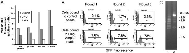
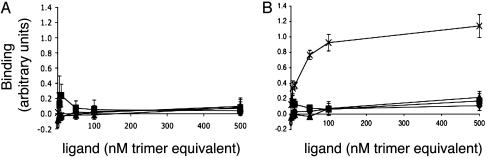
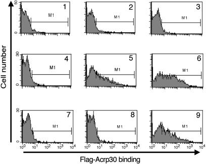
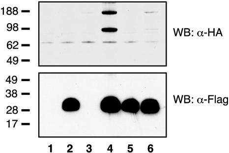
Acrp30/adiponectin is reduced in the serum of obese and diabetic individuals, and the genetic locus of adiponectin is linked to the metabolic syndrome. Recombinant adiponectin, administered to diet-induced obese mice, induced weight loss and improved insulin sensitivity. In muscle and liver, adiponectin stimulates AMP-activated protein kinase activation and fatty acid oxidation. To expression-clone molecules capable of binding adiponectin, we transduced a C2C12 myoblast cDNA retroviral expression library into Ba/F3 cells and panned infected cells on recombinant adiponectin linked to magnetic beads. We identified T-cadherin as a receptor for the hexameric and high-molecular-weight species of adiponectin but not for the trimeric or globular species. Only eukaryotically expressed adiponectin bound to T-cadherin, implying that posttranslational modifications of adiponectin are critical for binding. An adiponectin mutant lacking a conserved N-terminal cysteine residue required for formation of hexamer and high-molecular-weight species did not bind T-cadherin in coimmunoprecipitation studies. Although lacking known cellular functions, T-cadherin is expressed in endothelial and smooth muscle cells, where it is positioned to interact with adiponectin. Because T-cadherin is a glycosylphosphatidylinositol-anchored extracellular protein, it may act as a coreceptor for an as-yet-unidentified signaling receptor through which adiponectin transmits metabolic signals.


Figures

Fig. 1.
Fig. 1.
Cloning of T-cadherin as an adiponectin receptor. (A) FACS binding assay of tissue culture cells by using unpurified tissue culture supernatants containing Flag-tagged adiponectin. Adherent (C2C12, CHO) or suspension (Ba/F3) cells were incubated with control blocking solution or supernatants from mock vector-transfected HEK cells (left two columns) or unpurified cell supernatants containing 5′-or3′-FlagAcrp30 (right two columns; protein as depicted). Bound protein was detected by incubating cells with an APC-conjugated anti-Flag mAb and analyzed by FACS. Live cells were identified by exclusion of propidium iodide staining. The average of the median APC staining for each sample is shown; only C2C12 cells, and not CHO or Ba/F3 cells, demonstrate binding (n = 2). (B) FACS analysis of GFP expression of sequentially enriched pools of Ba/F3 cells transduced with a retroviral C2C12 cDNA expression library coexpressing GFP from an internal ribosome entry site. Naive Ba/F3 cells were infected with the cDNA library and enriched for binding to anti-Flag magnetic beads previously incubated with either 5′-Flag-Acrp30 or mock vector-transfected HEK cell supernatants. Three rounds of enrichment were performed as described in the text. After each round of binding and expansion of adherent cells, aliquots of cells were analyzed for GFP expression by FACS. After each round of sorting, the percentages of cells expressing GFP are indicated in the figure, compared with uninfected cells. (C) Genomic PCR amplification of integrated proviral cDNA insert of enriched Ba/F3 cell pools. Genomic DNA was prepared after the third round of enrichment from control bead-enriched Ba/F3 cells (lane 1; B Upper Right) or 5′-Flag-Acrp30 bead-enriched Ba/F3 cells (lane 2; B Lower Right). Primer pairs flanking the cDNA cloning site of the parental vector pBI-GFP were used to amplify the integrated proviral cDNA. A single 2.5-kb band was amplified from cells enriched for binding to 5′-Flag-Acrp30 beads, as shown by agarose gel electrophoresis and EtBr staining. Standards are indicated in kb.
Fig. 2.
Fig. 2.
ELISA-based binding assay. Flag-tagged recombinant proteins were incubated with CHO-GFP cells (A) or CHO-GFP-Tcad-expressing cells (B). Bound protein was detected by subsequent binding of anti-Flag Ab and labeled secondary Ab. ×, HEK-produced Hexamer/HMW 5′-Flag-Acrp30; ▴, HEK-produced trimeric 5′-Flag-Acrp30; ♦, bacterially produced globular adiponectin 3′-Flag-tagged; ▪, bacterially produced full-length adiponectin 3′-Flag-tagged. The concentration of added protein is plotted vs. the average binding signal (arbitrary units) ± SD (n = 4).
Fig. 3.
Fig. 3.
FACS-based binding assay. Cells (Ba/F3 or Ba/F3-GFP-Tcad) were incubated with or without Flag-tagged adiponectin; bound protein was detected by subsequent incubation with APC-labeled anti-Flag Ab and analyzed by FACS. Shown are histograms of APC fluorescence. Naive Ba/F3 cells (graphs 1–3) and Ba/F3-GFP-Tcad cells (graphs 4–9) were incubated without added protein (graphs 1 and 4), or 6 nM hexamer 5′-Flag-Acrp30 (graphs 2, 5, and 7–9) or 60 nM hexamer 5′-Flag-Acrp30 (graphs 3 and 6). The incubation reactions also included 60 nM hexameric untagged adiponectin (graph 7), 10 mM EDTA (graph 8), or a 20-fold excess of C1q by weight (graph 9).
Fig. 4.
Fig. 4.
Immunoprecipitation of cotransfected epitope-tagged T-cadherin and adiponectin. HEK cells were transfected with pcDNA alone (lane 1) or pcDNA expressing 5′-Flag-Acrp30 (lanes 2 and 4), 5′-Flag-C22A-Acrp30 (lanes 5 and 6), or HA-T-cadherin (lanes 3, 4, and 6). Forty-eight hours after transfection, aliquots of the Triton X-100 soluble cell lysates were immunoprecipitated on anti-Flag resin, electrophoresed on duplicate 4–12% polyacrylamide gels, and analyzed by immunobloting with anti-HA (Upper) or anti-Flag (Lower) mAbs conjugated to horseradish peroxidase. Standards are in kDa.

References

    1. Scherer, P. E., Williams, S., Fogliano, M., Baldini, G. & Lodish, H. F. (1995) J. Biol. Chem. 270, 26746–26749. - PubMed
    1. Tsao, T. S., Tomas, E., Murrey, H. E., Hug, C., Lee, D. H., Ruderman, N. B., Heuser, J. E. & Lodish, H. F. (2003) J. Biol. Chem. 278, 50810–50817. - PubMed
    1. Hara, K., Boutin, P., Mori, Y., Tobe, K., Dina, C., Yasuda, K., Yamauchi, T., Otabe, S., Okada, T., Eto, K., et al. (2002) Diabetes 51, 536–540. - PubMed
    1. Yamauchi, T., Kamon, J., Waki, H., Terauchi, Y., Kubota, N., Hara, K., Mori, Y., Ide, T., Murakami, K., Tsuboyama-Kasaoka, N., et al. (2001) Nat. Med. 7, 941–946. - PubMed
    1. Fruebis, J., Tsao, T. S., Javorschi, S., Ebbets-Reed, D., Erickson, M. R., Yen, F. T., Bihain, B. E. & Lodish, H. F. (2001) Proc. Natl. Acad. Sci. USA 98, 2005–2010. - PMC - PubMed

Publication types

MeSH terms