. 2004 Jun 29;101(26):9843-8.
doi: 10.1073/pnas.0403495101. Epub 2004 Jun 21.

The role of iron and copper molecules in the neuronal vulnerability of locus coeruleus and substantia nigra during aging

Affiliations

The role of iron and copper molecules in the neuronal vulnerability of locus coeruleus and substantia nigra during aging

Luigi Zecca et al. Proc Natl Acad Sci U S A. .

Abstract

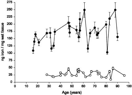
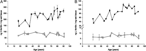
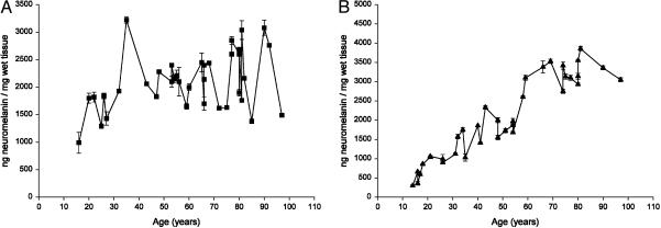
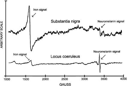
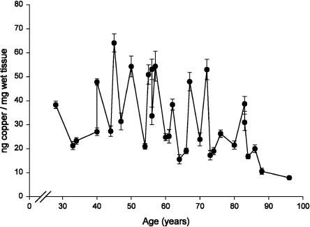
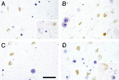
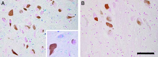
In this study, a comparative analysis of metal-related neuronal vulnerability was performed in two brainstem nuclei, the locus coeruleus (LC) and substantia nigra (SN), known targets of the etiological noxae in Parkinson's disease and related disorders. LC and SN pars compacta neurons both degenerate in Parkinson's disease and other Parkinsonisms; however, LC neurons are comparatively less affected and with a variable degree of involvement. In this study, iron, copper, and their major molecular forms like ferritins, ceruloplasmin, neuromelanin (NM), manganese-superoxide dismutase (SOD), and copper/zinc-SOD were measured in LC and SN of normal subjects at different ages. Iron content in LC was much lower than that in SN, and the ratio heavy-chain ferritin/iron in LC was higher than in the SN. The NM concentration was similar in LC and SN, but the iron content in NM of LC was much lower than SN. In both regions, heavy- and light-chain ferritins were present only in glia and were not detectable in neurons. These data suggest that in LC neurons, the iron mobilization and toxicity is lower than that in SN and is efficiently buffered by NM. The bigger damage occurring in SN could be related to the higher content of iron. Ferritins accomplish the same function of buffering iron in glial cells. Ceruloplasmin levels were similar in LC and SN, but copper was higher in LC. However, the copper content in NM of LC was higher than that of SN, indicating a higher copper mobilization in LC neurons. Manganese-SOD and copper/zinc-SOD had similar age trend in LC and SN. These results may explain at least one of the reasons underlying lower vulnerability of LC compared to SN in Parkinsonian syndromes.


Figures

Fig. 1.
Fig. 1.
Concentration of iron (ng/mg of wet tissue) in LC (○) and in SN (•)of human normal subjects during aging. Values are given as mean ± SEM (n = 2). In SN, data were best fit to a linear regression model (Y = 0.668X + 134.494, R2 = 0.158, P = 0.040).
Fig. 2.
Fig. 2.
Age trend of ferritins in SN and LC. (A) Concentration of H-ferritin (ng/mg of wet tissue) in LC (○) and in SN (•) of human normal subjects during aging. Values are expressed as mean ± SEM (n = 6). In SN, data were best fit to a linear regression model (Y = 1.151X + 159.455, R2 = 0.439, P = 0.002). (B) Concentration of L-ferritin (ng/mg of wet tissue) in LC (○) and in SN (•) of human normal subjects during aging. Values are expressed as mean ± SEM (n = 6). In SN, data were best fit to a linear regression model (Y = 0.886X + 33.830, R2 = 0.562, P = 0.0003).
Fig. 3.
Fig. 3.
Concentration of NM (ng/mg of wet tissue) in LC (A) and in SN (B) of human normal subjects during aging. Values are expressed as mean ± SEM (n = 2). Data were best fit to a linear regression model (Y = 8.345X + 1587.954, R2 = 0.127, P = 0.028 in LC; Y = 40.648X + 10.108, R2 = 0.872, P = 0.0001 in SN).
Fig. 4.
Fig. 4.
EPR spectra of NM-iron complex in SN and in LC.
Fig. 5.
Fig. 5.
Concentration of copper (ng/mg of wet tissue) in LC of human normal subjects during aging. Values are mean ± SEM (n = 2). Data were best fit to a linear regression model (Y = -0.331X + 51.935, R2 = 0.157, P = 0.025).
Fig. 6.
Fig. 6.
Cellular distribution of ferritins in SN and LC. Immunohistochemistry of H-ferritin in SN (A) and in LC (B). Immunohistochemistry of L-ferritin in SN (C) and in LC (D). Tissues are from a normal 83-year-old female subject. NM of dopaminergic neurons of SN and norepinephrine neurons of LC are brown granules. The H- and L-ferritin reaction products are violet. (Scale bar, 100 μm.) In SN, only a few neurons containing NM have a very light violet staining; the neurons then show a low content of H-ferritin. At higher magnification, a light violet staining is observed in a nonpigmented neuron (A). Many violet-staining oligodendrocytes occur in SN (A) and LC (B) with H-ferritin-positive reactions in nuclei and cytoplasm. L-ferritin staining is not detected in any NM neuron of both SN (C) and LC (D). At higher magnification in two nonpigmented neurons of LC, only a few spots of L-ferritin staining are present in the cytoplasm (D). Again, a high number of L-ferritin positive oligodendrocytes is observed in SN (C) and LC (D).
Fig. 7.
Fig. 7.
Iron histochemistry with modified Perls' staining of human SN (A) and LC (B) from a normal 88-year-old male subject. NM of dopaminergic neurons of SN and norepinephrine neurons of LC are brown granules, and iron deposits are colored in blue. (Scale bar, 100 μm.) NM-containing neurons in both SN (A) and LC (B) do not have the blue staining of iron. In SN (A), there are many iron-positive cells, which are most often oligodendrocytes with cytoplasmic iron deposits, and in LC (B) there are very few oligodendrocytes with light iron staining. Iron deposits are present also in the cytoplasm of nonpigmented neurons of SN (A) as shown at higher magnification. Iron deposits can be observed in the whole SN (A) parenchima with the exception of pigmented neurons, but they are completely absent in LC (B) parenchima.

References

    1. Foote, S. L., Freedman, R. & Oliver, A. P. (1975) Brain Res. 86, 229-242. - PubMed
    1. Arnsten, A. F. & Goldman-Rakic, P. S. (1987) J. Neural Transm. 24, 317-324. - PubMed
    1. Mason, S. T. & Fibiger, H. C. (1979) J. Comp. Neurol. 187, 703-724. - PubMed
    1. Nygren, L. G. & Olson, L. (1977) Brain Res. 132, 85-93. - PubMed
    1. German, D. C., Manaye, K. F., White, C. L. III, Woodward, D. J., McIntire, D. D., Smith, W. K., Kalaria, R. N. & Mann, D. M. (1992) Ann. Neurol. 32, 667-676. - PubMed

Publication types

MeSH terms