

doi: 10.1073/pnas.0308733101.
Epub 2004 Jun 21.
Relating microstructure to rheology of a bundled and cross-linked F-actin network in vitro
Affiliations
- PMID: 15210969
- PMCID: PMC470727
- DOI: 10.1073/pnas.0308733101
Item in Clipboard
Relating microstructure to rheology of a bundled and cross-linked F-actin network in vitro
Proc Natl Acad Sci U S A.
.
Abstract
The organization of individual actin filaments into higher-order structures is controlled by actin-binding proteins (ABPs). Although the biological significance of the ABPs is well documented, little is known about how bundling and cross-linking quantitatively affect the microstructure and mechanical properties of actin networks. Here we quantify the effect of the ABP scruin on actin networks by using imaging techniques, cosedimentation assays, multiparticle tracking, and bulk rheology. We show how the structure of the actin network is modified as the scruin concentration is varied, and we correlate these structural changes to variations in the resultant network elasticity.
Figures
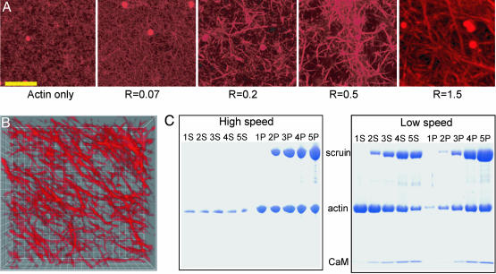
Changes in the degree of bundling at various R values. (A) Confocal images of an F-actin:scruin network at various R values. The rightmost image is an assembled 3D projection of 50 images with 100-nm intervals. (Scale bar, 10 μm.) (B) Three-dimensional deconvolved image of a 1:2 (scruin:actin) network. Each grid measures 1 μm. (C) Scanned image of a SDS/polyacrylamide gel. The sample numbers, 1, 2, 3, 4, and 5, correspond to R = 0, 0.07, 0.2, 0.5, and 1, respectively, at a fixed cA = 11.9 μM. S, supernatant after centrifugation; P, pellet after centrifugation. High-speed cosedimentation assay data shows that the F-actin density is unaffected by the presence of scruin and that all of scruin binds to F-actin. Low-speed assay data shows the degree of bundling; although all scruin binds to F-actin, not all of the scruin-decorated F-actins assemble into thick bundles. CaM, calmodulin.
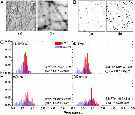
Characterization of the bundle thickness, DB, and pore size, ξ, distribution at various R values. (A) EM images of the actin-only (a) and R = 1(b) samples. (Scale bar, 200 nm.) (B) Two-dimensional map of the particle trajectories to demonstrate ξ and the degree of heterogeneity at R = 0.03 (a) and R = 1(b). (Scale bar, 1 μm.) (C) The distribution of pore sizes at various R values for cA = 11.9 μM measured with MPT (solid red) and confocal microscopy (CF, striated blue). We observed both the mean and the width of the distribution of ξ increasing as R increased. P(ξ), normalized probability.
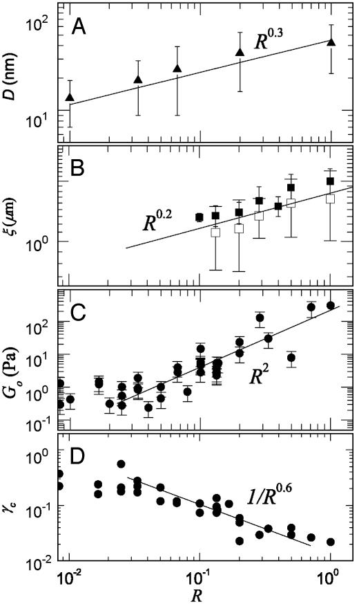
The bundle thickness, mesh size, elastic modulus, and critical strain as a function of R at cA = 11.9 μM. (A) The average DB at various R values was measured from the digitized EM images and shows DB ∼ R0.3. A single actin filament is ≈7 nm in diameter, and DB becomes as large as 65 nm at R = 1. (B) ξ is measured by using both MPT (▪) and confocal imaging (□). Results show that ξ at R = 0.1 is 2-fold larger than that predicted for an entangled actin network and follows the scaling of ξ ∼ R0.2. (C) Go was measured by using bulk rheology, and the best fit for the data follows Go ∼ R2 (solid line). (D) The strain at which we observe the onset of nonlinearity, γcrit, of the actin–scruin composite networks at various R values, showing a scaling of γcrit ∼ R-0.6.
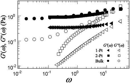
G′ (ω) (solid symbols) and G″ (ω) (open symbols) at cA = 11.9 μM and R = 0.03 measured with 1-P and 2-P microrheology and bulk rheology. Although the elastic moduli, Go, measured with both 1-P and 2-P microrheology with BSA-coated particles match well with the bulk measurement, 2-P microrheology shows an excellent agreement with the bulk rheology.
References
-
- Kreis, T. & Vale, R., eds. (1999) Guidebook to the Cytoskeletal and Motor Proteins (Oxford Univ. Press, New York), 2nd. Ed.
-
- Cohan, C. S., Welnhofer, E. A., Zhao, L., Matsumura, F. & Yamashiro, S. (2001) Cell Motil. Cytoskeleton 48, 109-120. - PubMed
-
- Fabry, B., Maksym, G. N., Butler, J. P., Glogauer, M., Navajas, D. & Fredberg, J. J. (2001) Phys. Rev. Lett. 87, 148102-148106. - PubMed
Publication types
MeSH terms
Substances
Grants and funding
LinkOut - more resources
Full Text Sources
