. 2004 Jun 29;101(26):9716-21.
doi: 10.1073/pnas.0305659101. Epub 2004 Jun 21.

The louse-borne human pathogen Bartonella quintana is a genomic derivative of the zoonotic agent Bartonella henselae

Affiliations

The louse-borne human pathogen Bartonella quintana is a genomic derivative of the zoonotic agent Bartonella henselae

Cecilia M Alsmark et al. Proc Natl Acad Sci U S A. .

Abstract

We present the complete genomes of two human pathogens, Bartonella quintana (1,581,384 bp) and Bartonella henselae (1,931,047 bp). The two pathogens maintain several similarities in being transmitted by insect vectors, using mammalian reservoirs, infecting similar cell types (endothelial cells and erythrocytes) and causing vasculoproliferative changes in immunocompromised hosts. A primary difference between the two pathogens is their reservoir ecology. Whereas B. quintana is a specialist, using only the human as a reservoir, B. henselae is more promiscuous and is frequently isolated from both cats and humans. Genome comparison elucidated a high degree of overall similarity with major differences being B. henselae specific genomic islands coding for filamentous hemagglutinin, and evidence of extensive genome reduction in B. quintana, reminiscent of that found in Rickettsia prowazekii. Both genomes are reduced versions of chromosome I from the highly related pathogen Brucella melitensis. Flanked by two rRNA operons is a segment with similarity to genes located on chromosome II of B. melitensis, suggesting that it was acquired by integration of megareplicon DNA in a common ancestor of the two Bartonella species. Comparisons of the vector-host ecology of these organisms suggest that the utilization of host-restricted vectors is associated with accelerated rates of genome degradation and may explain why human pathogens transmitted by specialist vectors are outnumbered by zoonotic agents, which use vectors of broad host ranges.


Figures

Fig. 1.
Fig. 1.
The B. henselae and the B. quintana genome maps and the location of genes with homologs in other α-proteobacterial genomes. The outer circle shows predicted coding regions on the plus strand in B. henselae color-coded by role categories. The second circle shows predicted coding regions on the minus strand color-coded by role categories. Third circle, genes with orthologs in B. quintana. Fourth circle, top hits to B. melitensis according to replicon: blue, main chromosome (I); red, chromosome II. Fifth circle, top hits to A. tumefaciens according to replicon: blue, circular chromosome; red, linear chromosome, green, plasmid pAtC58, dark green, plasmid pTiC58. Sixth circle, top hits to Mesorhizobium loti according to replicon: blue, main chromosome; red, plasmid pMLa; green, plasmid pMLb. Seventh circle, top hits to S. meliloti according to replicon: blue, main chromosome; red, plasmid pSymA; green, plasmid pSymB. Eighth circle, B. henselae islands and islets in gray, Bartonella islands in orange. Ninth circle, tRNAs in green, rRNAs in red. Tenth circle, integrase remnants in pink. Eleventh circle, pseudogenes in blue and extensively degraded gene remnants in light blue. Twelfth circle, repeats, the length of the line is proportional to the length of the repeated region, and the color gradient represents percent similarity ranging from 100% (red) to 75% (yellow). Thirteenth circle, GC skew in sliding window of 15 kb, step size 7 kb. Innermost circle, deviation from average GC content in sliding windows of 15 kb, step size 7 kb. Numbers refer to prophage (1), B. henselae specific islands of 72 kb (2), 9 kb (3), 34 kb (4), and Bartonella specific island (5).
Fig. 2.
Fig. 2.
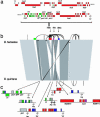
Schematic illustration of the rearrangements observed between B. henselae and B. quintana. (a) Gene order structure in the 72-kb island of B. henselae. (b) The breakpoints for rearrangements coincide with species-specific islands and repeated sequences. B. henselae-specific islands are color-coded (green, the prophage; red, the 72-kb island; purple, the 9-kb island; blue, the 34-kb island). Repeated regions longer than 1,000 bp are drawn as arcs between the two repeat copies. Black horizontal bars represent gene-order synteny with B. melitensis. (c) Pseudogenes and remnants of genes in these islands at the B. quintana breakpoints are marked with squares in the corresponding colors. Pseudogenes other than phage and integrase are shown in gray color.
Fig. 3.
Fig. 3.
Pseudogenes. The number of pseudogenes in B. henselae (open bars) and B. quintana (filled bars) is shown for different classes of genes.
Fig. 4.
Fig. 4.
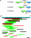
Integrase genes. (a) Size of the spacer region flanked by ubiA and phdX in B. henselae, B. quintana, and B. melitensis.(b) Size of integrase gene remnants in B. henselae sorted according to length. Sequence similarity against the full-length integrase gene was inferred by using the tBLASTn algorithm (black, E < 1e-30; red, E < 1e-20; green, E < 1e-4).

References

    1. Koehler, J. E. (1996) Adv. Pediatr. Infect. Dis. 11, 1-27. - PubMed
    1. Regnery, R. L., Anderson, B. E., Clarridge, J. E., III, Rodriguez-Barradas, M. C., Jones, D. C. & Carr, J. H. (1992) J. Clin. Microbiol. 30, 265-274. - PMC - PubMed
    1. Anderson, B. E. & Neuman, M. A. (1997) Clin. Microbiol. Rev. 10, 203-219. - PMC - PubMed
    1. Brouqui, P. & Raoult, D. (1996) Res. Microbiol. 147, 719-731. - PubMed
    1. Dehio, C., Meyer, M., Berger, J., Schwarz, H. & Lanz, C. (1997) J. Cell Sci. 110, 2141-2151. - PubMed

Publication types

MeSH terms

Associated data