

doi: 10.1073/pnas.0402700101.
Epub 2004 Jun 21.
Peptide and protein sequence analysis by electron transfer dissociation mass spectrometry
Affiliations
- PMID: 15210983
- PMCID: PMC470779
- DOI: 10.1073/pnas.0402700101
Item in Clipboard
Peptide and protein sequence analysis by electron transfer dissociation mass spectrometry
Proc Natl Acad Sci U S A.
.
Abstract
Peptide sequence analysis using a combination of gas-phase ion/ion chemistry and tandem mass spectrometry (MS/MS) is demonstrated. Singly charged anthracene anions transfer an electron to multiply protonated peptides in a radio frequency quadrupole linear ion trap (QLT) and induce fragmentation of the peptide backbone along pathways that are analogous to those observed in electron capture dissociation. Modifications to the QLT that enable this ion/ion chemistry are presented, and automated acquisition of high-quality, single-scan electron transfer dissociation MS/MS spectra of phosphopeptides separated by nanoflow HPLC is described.
Figures
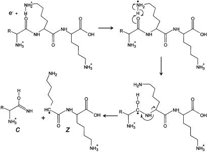
Fragmentation scheme for production of c- and z-type ions after reaction of a low-energy electron with a multiply protonated peptide.

Fragmentation scheme for production of b- and y-type ions by CAD of a multiply protonated peptide.
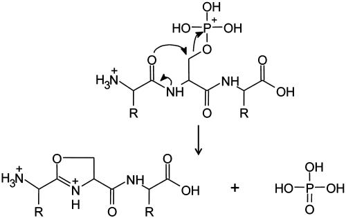
Fragmentation scheme for loss of phosphoric acid from a multiply protonated phosphopeptide by CAD.
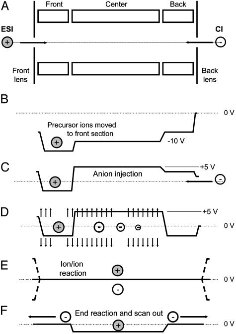
Schematic of steps involved in the operation of the LTQ mass spectrometer for peptide sequence analysis by ETD. (A) Injection of multiply protonated peptide molecules (precursor ions) generated by ESI. (B) Application of a dc offset to move the precursor ions to the front section of the linear trap. (C) Injection of negatively charged reagent ions from the CI source into the center section of the linear trap. (D) Application of a supplementary dipolar broadband ac field to eject all ions except those within 3 mass-unit windows centered around the positively charged precursor ions and the negatively charged electron-donor reagent ions. (E) Removal of the dc potential well and application of a secondary RF voltage (100 V zero to peak, 600 kHz) to the end lens plates of the linear trap to allow positive and negative ion populations to mix and react. (F) Termination of ion/ion reactions by axial ejection of negatively charged reagent ions while retaining positive ions in the center section of the trap. This is followed by mass-selective, radial ejection of positively charged fragment ions to record the resulting MS/MS spectrum.

Single-scan ETD MS/MS spectrum resulting from a 50-msec reaction of the triply charged phosphopeptide, LPISASHpSpSKTR, at m/z 482, with anthracene anions. Predicted m/z values for fragment ions of types c and z are shown above and below the sequence, respectively. Those observed are underlined. Note that both z5 and c7 have m/z values that overlap with the ion cluster containing the product of proton abstraction, the (M + 2H)+2 ion at m/z 722. All other possible ions of types c and z appear in the spectrum. The total experiment time was ≈300 msec.

Data-dependent analysis of a peptide mixture by using a combination of nHPLC-μESI and ETD-MS/MS. (A) Total ion chromatogram (peaks are ≈10 sec wide). (B) Single-scan, 500- to 600-msec, ETD spectrum recorded on 100 fmol of the triply protonated peptide, DRVYIHPFHL. (C) Single-scan, 500- to 600-msec, ETD spectrum recorded on 1 fmol of the triply protonated peptide, DRpSPIRGpSPR.

Comparison of single-scan (500- to 600-msec) CAD and ETD mass spectra recorded during data-dependent analyses (nHPLC-μESI-MS/MS) of phosphopeptides generated in a tryptic digest of human nuclear proteins. All peptides were converted to methyl esters and subjected to immobilized metal affinity chromatography before analysis by MS. (A) CAD spectrum dominated by fragment ions corresponding to the loss of phosphoric acid and either methanol or water. (B) ETD spectrum containing 13 of 14 possible c- and z-type product ions. Note that the spectrum is devoid of fragment ions corresponding to the loss of phosphoric acid.
References
-
- Zubarev, R. A., Kelleher, N. L. & McLafferty, F. W. (1998) J. Am. Chem. Soc. 120, 3265-3266.
-
- Zubarev, R. A., Kruger, N. A., Fridriksson, E. K., Lewis, M. A., Horn, D. M., Carpenter, B. K. & McLafferty, F. W. (1999) J. Am. Chem. Soc. 121, 2857-2862.
-
- Zubarev, R. A. (2003) Mass Spectrom. Rev. 22, 57-77. - PubMed
-
- Cerda, B. A., Horn, D. M., Breuker, K., Carpenter, B. K. & McLafferty, F. W. (1999) Eur. J. Mass Spectrom. (Chichester, England) 5, 335-338.
-
- Zubarev, R. A., Haselmann, K. F., Budnik, B., Kjeldsen, F. & Jensen, F. (2002) Eur. J. Mass Spectrom. (Chichester, England) 8, 337-349.
Publication types
MeSH terms
Substances
Grants and funding
LinkOut - more resources
Full Text Sources
Other Literature Sources
