. 2004 Nov;92(5):2920-32.
doi: 10.1152/jn.00064.2004. Epub 2004 Jun 22.

Neuronal activation in the medulla oblongata during selective elicitation of the laryngeal adductor response

Affiliations

Neuronal activation in the medulla oblongata during selective elicitation of the laryngeal adductor response

Ranjinidevi Ambalavanar et al. J Neurophysiol. 2004 Nov.

Abstract

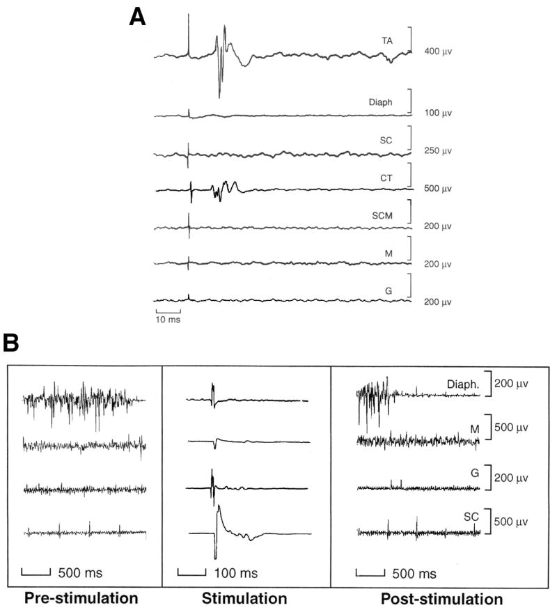
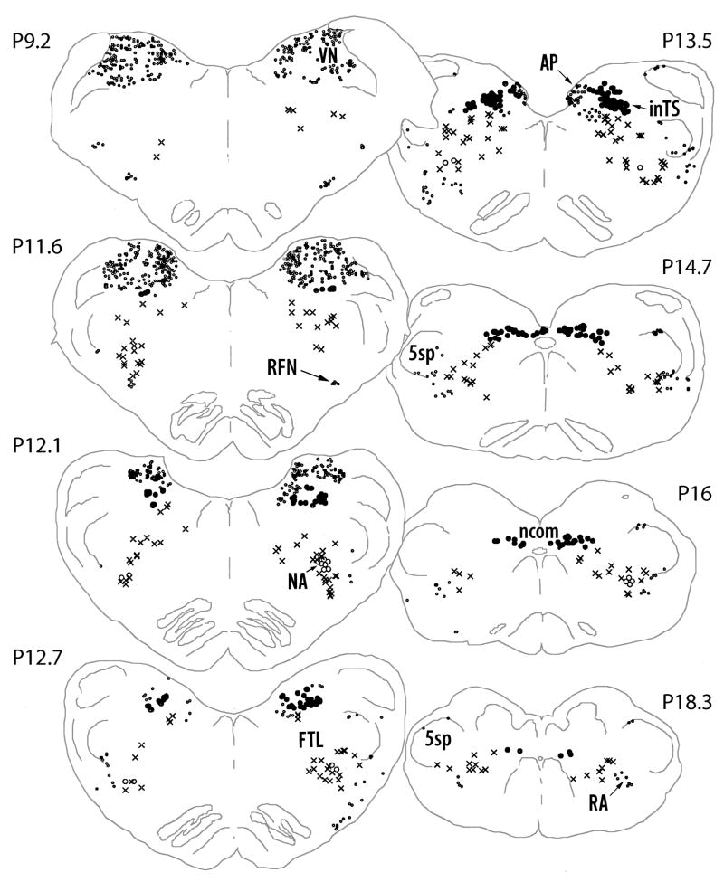
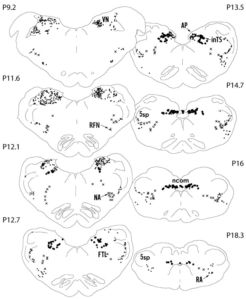
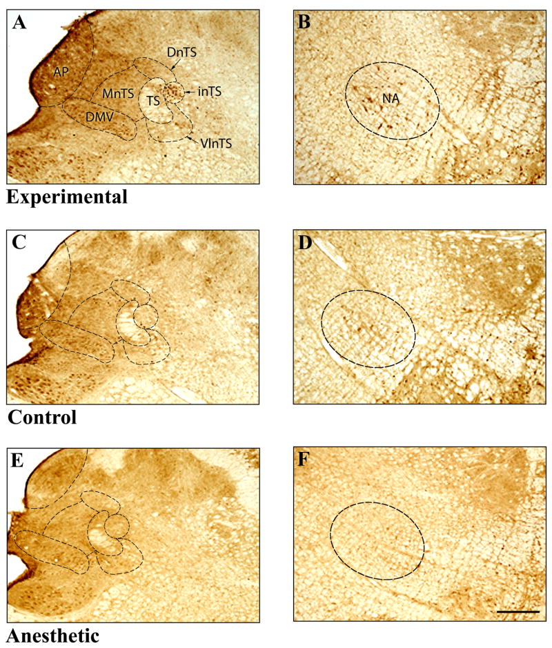
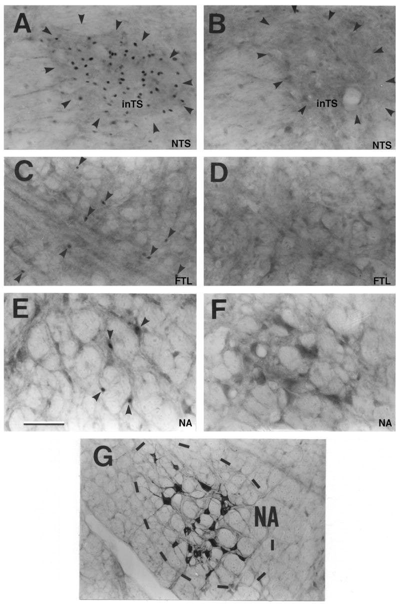
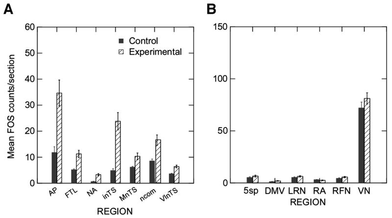
Swallow and cough are complex motor patterns elicited by rapid and intense electrical stimulation of the internal branch of the superior laryngeal nerve (ISLN). The laryngeal adductor response (LAR) includes only a laryngeal response, is elicited by single stimuli to the ISLN, and is thought to represent the brain stem pathway involved in laryngospasm. To identify which regions in the medulla are activated during elicitation of the LAR alone, single electrical stimuli were presented once every 2 s to the ISLN. Two groups of five cats each were studied; an experimental group with unilateral ISLN stimulation at 0.5 Hz and a surgical control group. Three additional cats were studied to evaluate whether other oral, pharyngeal, or respiratory muscles were activated during ISLN stimulation eliciting LAR. We quantified < or = 22 sections for each of 14 structures in the medulla to determine if regions had increased Fos-like immunoreactive neurons in the experimental group. Significant increases (P < 0.0033) occurred with unilateral ISLN stimulation in the interstitial subnucleus, the ventrolateral subnucleus, the commissural subnucleus of the nucleus tractus solitarius, the lateral tegmental field of the reticular formation, the area postrema, and the nucleus ambiguus. Neither the dorsal motor nucleus of the vagus, usually active for swallow, nor the nucleus retroambiguus, retrofacial nucleus, and the lateral reticular nucleus, usually active for cough, were active with elicitation of the laryngeal adductor response alone. The results demonstrate that the laryngeal adductor pathway is contained within the broader pathways for cough and swallow in the medulla.


Figures

Figure 1
Figure 1
A. The average of over 114 responses to stimulation of the internal branch of the superior laryngeal at 2 s intervals (0.5 Hz) showing active responses only in the laryngeal muscles, the thyroarytenoid (TA) and the cricothyroid (CT) muscles at 10 ms on the same side as the stimulation. Simultaneous recordings from the diaphragm (Diaph), the superior constrictor of the pharyngeal wall (SC), the sternocleidomastoid (SCM), the masseter (M) and the genioglossus (G) on the same side show the absence of responses in muscles involved in swallow and cough. B. Examples of recordings of respiratory and swallowing muscles (Diaph.=diaphragm, M= masseter, G= genioglossus, and SC=superior constrictor) 2 s before (pre-stimulation), during the elicitation of the laryngeal adductor response (Stimulation) and 2 s after (Post-stimulation) to demonstrate that changes in muscle tone after with stimulation in comparison with before stimulation. Diaphragmatic activity is the phasic activity increasing with inspiration. The Stimulation panel shows averages of muscles responses to 114 stimulation trials at 0.5 Hz with stimulation artifact and no clear muscle response in contrast with the thyroarytenoid (TA) and cricothyroid (CT) muscle recordings in A. The spontaneous muscle activity is shown before and after stimulation for the diaphragm (Diaph), masseter (M), superior constrictor (SC) and genioglossus (G). None of these muscles show changes in background activity following ISLN stimulation.
Figure 2
Figure 2
Computer video drawings of representative sections through the medulla oblongata from a cat that had ISLN stimulation). The approximate rostrocaudal levels of the Berman’s atlas coordinates are marked on the figure (Berman 1968). AP - area postrema; S; FTL - lateral tegmental field of the reticular formation; inTS - interstitial subnucleus of the NTS;; ncom - commisural subnucleus of the NTS; NA - nucleus ambiguous; RA - retroambigual nucleus; RFN - retrofacial nucleus; VN - inferior vestibular nucleus;; 5sp - spinal trigeminal nucleus.
Figure 3
Figure 3
Computer video drawings of representative sections through the medulla oblongata from an unstimulated cat (Control). The approximate rostrocaudal levels of the Berman’s atlas coordinates are marked on the figure (Berman 1968). AP - area postrema;; FTL - lateral tegmental field of the reticular formation; inTS - interstitial subnucleus of the NTS;; ncom - commisural subnucleus of the NTS; NA - nucleus ambiguous; RA - retroambigual nucleus; RFN - retrofacial nucleus; VN - inferior vestibular nucleus; ; 5sp - spinal trigeminal nucleus.
Figure 4
Figure 4
Photomicrographs of a section through the brain stem at the level of the area postrema from an experimental (A, B), surgical control (C,D) and an anesthetic control (E,F) cat showing FLI neurons in different subnuclei of the NTS (A,C,E) and in the NA (B,D,F). Approximate boundaries of the subnuclei are shown by dotted lines. AP-area postrema, DMV – dorsal motor nucleus of the vagus, DnTS – dorsal subnucleus of the nucleus tractus solitarii (NTS),, NA - nucleus ambiguus, inTS - interstitial subnucleus of the NTS, MnTS medial subnucleus of the NTS, TS – solitary tract, VlnTS - ventrolateral subnucleus of the NTS.. Scale bar = 500μm.
Figure 5
Figure 5
Fos-like immunoreactivity in the inTS (A, B), FTL (C, D) and the NA (E,F). Many FLI neurons can be seen in all three brain stem regions of the stimulated than the nonstimulated control cats. Laryngeal motor neurons retrogradely labeled in the NA are shown in G. (Scale bar = 100 μm).
Figure 6
Figure 6
Mean and standard errors in the number of FLI neurons counted for each structure in the control (Control) and the experimental (Experimental) groups. A provides the plots of the structures where the two groups differed significantly (p<.0033) or showed a trend towards a difference (MnTS, p=.008). Structures are AP= area postrema, FTL=lateral tegmental field, NA= nucleus ambiguus, inTS=interstitial subnucleus of the nucleus tractus solitarii (NTS), ncom= commissural subnucleus of the NTS, and the VlnTS= ventrolateral subnucleus of the NTS. B provides plots of the other structures which did not differ between the two groups in the number of FLI neurons. Structures are 5sp=spinal trigeminal nucleus, DMV= dorsal motor nucleus of the vagus, LRN= lateral reticular nucleus, RA= retroambiguus, RFN= retrofacial nucleus, and VN=vestibular nucleus.

Similar articles

Cited by

References

    1. Abu-Shaweesh JM, Dreshaj IA, Haxhiu MA, Martin RJ. Central GABAergic mechanisms are involved in apnea induced by SLN stimulation in piglets. J Appl Physiol. 2001;90:1570–1576. - PubMed
    1. Altschuler SM, Bao XM, Bieger D, Hopkins DA, Miselis RR. Viscerotopic representation of the upper alimentary tract in the rat: sensory ganglia and nuclei of the solitary and spinal trigeminal tracts. J Comp Neurol. 1989;283:248–268. - PubMed
    1. Ambalavanar R, Ludlow CL, Wenthold RJ, Tanaka Y, Damirjian M, Petralia RS. Glutamate receptor subunits in the nucleus of the tractus solitarius and other regions of the medulla oblongata in the cat. J Comp Neurol. 1998;402:75–92. - PubMed
    1. Ambalavanar R, Purcell L, Miranda M, Evans F, Ludlow CL. Selective suppression of late laryngeal adductor responses by N-Methyl-D-Asparate receptor blockade in the cat. J Neurophysiol. 2002;87:1252–1262. - PubMed
    1. Ambalavanar R, Tanaka Y, Damirjian M, Ludlow CL. Laryngeal afferent stimulation enhances fos immunoreactivity in periaqueductal gray in the cat. J Comp Neurology. 1999;409:411–423. - PubMed

Publication types

LinkOut - more resources