. 2004 Jul;72(7):3863-8.
doi: 10.1128/IAI.72.7.3863-3868.2004.

Cleavage of host keratin 8 by a Chlamydia-secreted protease

Affiliations

Cleavage of host keratin 8 by a Chlamydia-secreted protease

Feng Dong et al. Infect Immun. 2004 Jul.

Abstract

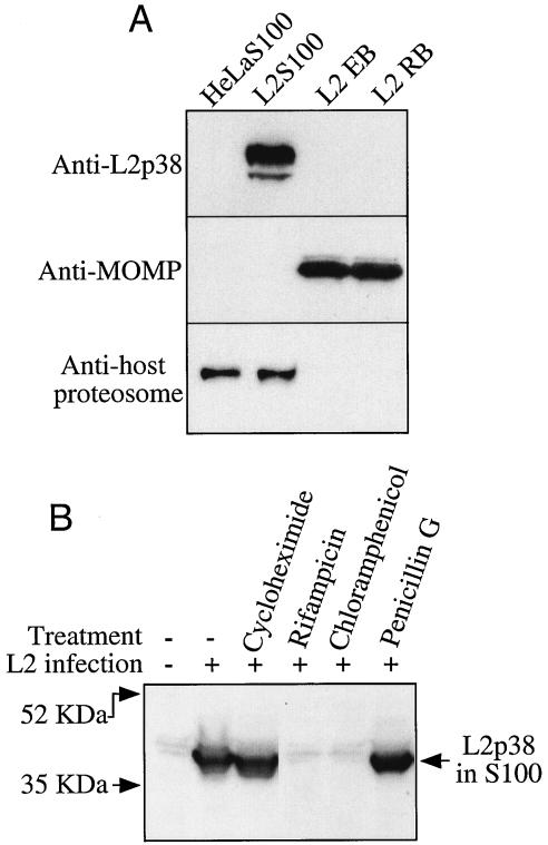
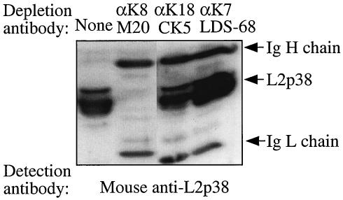
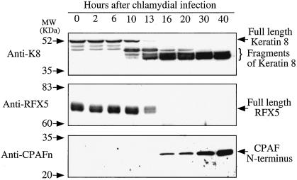
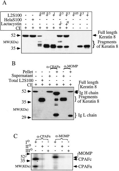
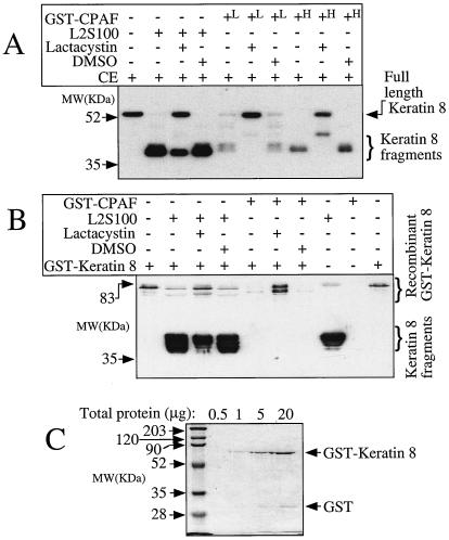
Chlamydiae have to replicate within a cytoplasmic vacuole in eukaryotic cells. Expansion of the chlamydia-laden vacuole is essential for chlamydial intravacuolar replication, which inevitably causes host cell cytoskeleton rearrangements. A cleavage fragment of keratin 8 corresponding to the central rod region was detected in the soluble fraction of chlamydia-infected cells. Since keratin 8 is a major component of the intermediate filaments in simple epithelial cells, cleavage of keratin 8 may increase the solubility of the host cell cytoskeleton and thus permit vacuole expansion in chlamydia-infected cells. A chlamydia-secreted protease designated CPAF (chlamydial protease/proteasome-like activity factor) was both necessary and sufficient for keratin 8 cleavage in chlamydia-infected cells, suggesting that chlamydiae have evolved specific mechanisms for modifying the host cell cytoskeleton.


Figures

FIG. 1.
FIG. 1.
Identification of a unique protein designated L2p38 in the cytosol of chlamydia-infected cells. (A) Anti-L2p38 antibody was used to detect L2p38 in the cytosol of HeLa cells that were not infected (HeLaS100) or were infected with C. trachomatis serovar L2 (L2S100) and in the EBs (L2 EB) or RBs (L2 RB) of purified L2 organisms on a Western blot. L2p38 was detected only in L2S100 (top panel). Parallel samples were also used for detection of MOMP (monoclonal antibody MC22) (middle panel) and a host 20S proteosomal subunit (bottom panel). (B) L2-infected HeLa cells were treated with the antibiotics indicated at the top, and the cytosol fractions (S100) derived from the culture samples were subjected to a Western blot analysis with the anti-L2p38 antibody. Both rifampin (final concentration, 1 μg/ml) and chloramphenicol (60 μg/ml), but not penicillin G (100 U/ml) or cycloheximide (2 μg/ml), blocked generation of L2p38. All antibiotics were added at the beginning of the infection, and the concentrations were maintained throughout the infection.
FIG. 2.
FIG. 2.
L2p38 represents the central rod domain of host cell keratin 8. Mouse monoclonal antibodies against human keratin 8 (αK8 M20), keratin 18 (αK18 CK5), and keratin 7 (αK7 LDS-68) were used to precipitate L2p38 in the cytosol of chlamydia-infected HeLa cells (L2S100). The precipitation procedure was repeated once in order to completely deplete the corresponding antigens in the supernatants. The final remaining supernatants were subjected to Western blot analysis with the anti-L2p38 antibody. Note that only the M20 antibody recognizing an epitope in the keratin 8 rod region depleted L2p38 from the L2S100 cytosol sample, confirming that L2p38 contains the keratin 8 rod region. Ig H chain, immunoglobulin heavy chain; Ig L chain, immunoglobulin light chain.
FIG. 3.
FIG. 3.
Correlation of keratin 8 cleavage with degradation of RFX5 (as a measure of CPAF activity) and expression of CPAF during chlamydial infection. At various times after chlamydial infection, culture samples were extracted with an NP-40 buffer and used for Western blot analysis with antibody against keratin 8 (M20) to monitor keratin 8 cleavage (top panel), with antibody against RFX5 (rabbit anti-RFX5) to assess CPAF activity (middle panel), and with antibody against CPAF (mouse anti-CPAF N-terminal region [Anti-CPAFn]) to evaluate CPAF expression (bottom panel). Note that keratin 8 cleavage correlated well with both RFX5 degradation and CPAF expression. Also note that when HeLa cells were extracted with the NP-40 buffer, full-length keratin 8 with a molecular mass of 52 kDa appeared in the soluble fraction (top panel). However, when HeLa cells were subjected to Dounce homogenization to make S100, no full-length keratin 8 was detected in the supernatant (Fig. 1A, top panel). MW, molecular mass.
FIG. 4.
FIG. 4.
CPAF is necessary for keratin 8 cleavage in the cytosol of chlamydia-infected cells. (A) A cell-free cleavage assay was used to evaluate the ability of L2S100 or HeLaS100 to cleave keratin 8 in CE. L2S100 was used at three different concentrations (equivalent to an L2S100 stock volume of 0.05, 0.5, or 1 μl). HeLaS100 was similarly used at a concentration of 5 μl per reaction mixture. Lactacystin was used at a final concentration of either 20 μM or 100 μM. Note that the keratin 8 fragment in L2S100 alone was detectable in 0.5- and 1-μl samples but not in 0.05-μl samples. (B) An antibody depletion approach was used to evaluate whether CPAF is necessary for cleavage of keratin 8. Monoclonal antibodies against either MOMP or CPAF were used to precipitate corresponding antigens in the cytosol extracts of chlamydia-infected cells (L2S100). To completely deplete the corresponding antigens from L2S100, the precipitation procedure was repeated once. The pellets from the first precipitation and the final supernatants were both evaluated for the ability to cleave keratin 8 in the cell-free cleavage assay, as described above. Ig H chain, immunoglobulin heavy chain; Ig L chain, immunoglobulin light chain. (C) The antibody depletion efficiency was monitored by using radiolabeled cell lysates. Both anti-CPAFn antibody (monoclonal antibody 54b) and anti-MOMP antibody (monoclonal antibody MC22) successfully precipitated the corresponding antigens during the first precipitation (Io). However, in the second precipitation (IIo) minimal amounts of the antigens were precipitated, and after the third precipitation (IIIo) the preparation did not contain detectable amounts of the antigens, indicating that the first two precipitations completely removed the antigens from the cell lysates. The anti-CPAFn antibody 54b is known to precipitate both the N-terminal (CPAFn) and C-terminal (CPAFc) fragments of CPAF (29). MW, molecular mass.
FIG. 5.
FIG. 5.
CPAF is sufficient for cleavage of keratin 8. (A) Purified recombinant GST-CPAF was used to cleave keratin 8 in CE in a cell-free assay, and L2S100 was used as a positive control for monitoring the quality of the assay. Keratin 8 and its cleavage fragments were monitored by using the M20 antibody in a Western blot assay. GST-CPAF successfully cleaved keratin 8 at a final concentration of either 0.2 μM (L) or 0.6 μM (H), and the cleavage was inhibited by 50 μM lactacystin but not by the solvent DMSO alone. (B) Cell-free assay similar to the assay whose results are shown in panel A, except that purified recombinant GST-keratin 8 (free of other mammalian component contamination) was used as the substrate. Both GST-CPAF and L2S100 cleaved the recombinant keratin 8 and lactacystin, and DMSO did not inhibit the cleavage. GST-CPAF was used at a concentration of 0.2 μM, L2S100 was used at a concentration of at 0.5 μl/reaction mixture, lactacystin was used at a concentration of 50 μM, and GST-keratin 8 was used at a concentration of 1 μg/reaction mixture. It is not clear why no degradation fragment was detected in the sample in which GST-keratin 8 was cleaved by GST-CPAF. (C) Purified recombinant GST-keratin 8 with a molecular mass of 78 kDa was analyzed on an SDS-polyacrylamide gel stained with Coomassie blue dye. Although free GST was detected in lanes loaded with 5 to 20 μg of protein, we used 1 μg of protein as the substrate in panel B. MW, molecular mass.

References

    1. Askienazy-Elbhar, M., and J. H. Suchet. 1999. Persistent “silent” Chlamydia trachomatis female genital tract infections. Infect. Dis. Obstet. Gynecol. 7:31-34. - PMC - PubMed
    1. Beatty, W. L., R. P. Morrison, and G. I. Byrne. 1994. Persistent chlamydiae: from cell culture to a paradigm for chlamydial pathogenesis. Microbiol. Rev. 58:686-699. - PMC - PubMed
    1. Casanova, L., A. Bravo, F. Were, A. Ramirez, J. J. Jorcano, and M. Vidal. 1995. Tissue-specific and efficient expression of the human simple epithelial keratin 8 gene in transgenic mice. J. Cell Sci. 108:811-820. - PubMed
    1. Chen, P. H., D. A. Ornelles, and T. Shenk. 1993. The adenovirus L3 23-kilodalton proteinase cleaves the amino-terminal head domain from cytokeratin 18 and disrupts the cytokeratin network of HeLa cells. J. Virol. 67:3507-3514. - PMC - PubMed
    1. Ditzel, H. J., M. C. Strik, M. K. Larsen, A. C. Willis, A. Waseem, K. Kejling, and J. C. Jensenius. 2002. Cancer-associated cleavage of cytokeratin 8/18 heterotypic complexes exposes a neoepitope in human adenocarcinomas. J. Biol. Chem. 277:21712-21722. - PubMed

Publication types