. 2004 Jul;72(7):3890-901.
doi: 10.1128/IAI.72.7.3890-3901.2004.

Identification and characterization of a novel uropathogenic Escherichia coli-associated fimbrial gene cluster

Affiliations

Identification and characterization of a novel uropathogenic Escherichia coli-associated fimbrial gene cluster

Eric L Buckles et al. Infect Immun. 2004 Jul.

Abstract

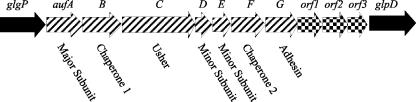
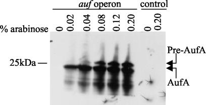
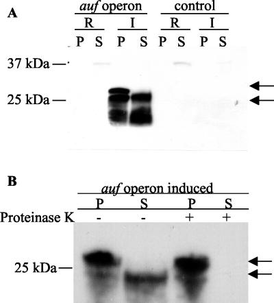
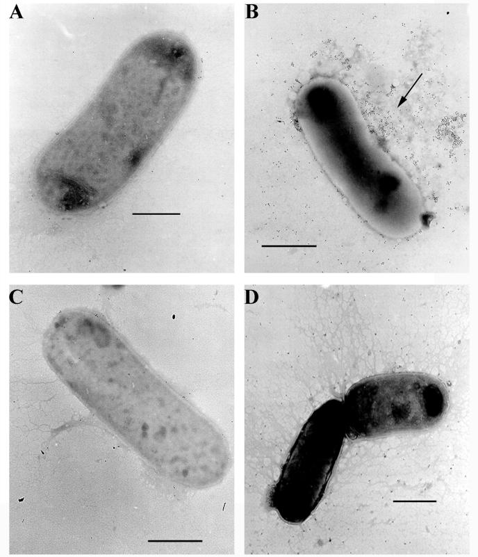
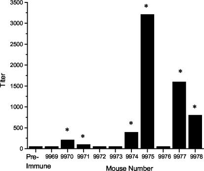
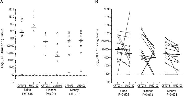
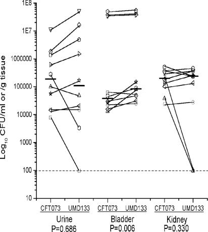
Recently, we identified a fimbrial usher gene in uropathogenic Escherichia coli strain CFT073 that is absent from an E. coli laboratory strain. Analysis of the CFT073 genome indicates that this fimbrial usher gene is part of a novel fimbrial gene cluster, aufABCDEFG. Analysis of a collection of pathogenic and commensal strains of E. coli and related species revealed that the auf gene cluster was significantly associated with uropathogenic E. coli isolates. For in vitro expression analysis of the auf gene cluster, RNA was isolated from CFT073 bacteria grown to the exponential or stationary phase in Luria-Bertani broth and reverse transcriptase PCR (RT-PCR) with oligonucleotide primers specific to the major subunit, aufA, was performed. We found that aufA is expressed in CFT073 only during the exponential growth phase; however, no expression of AufA protein was observed by Western blotting, indicating that under these conditions, the expression of the auf gene cluster is low. To determine if the auf gene cluster is expressed in vivo, RT-PCR was performed on bacteria from urine samples of mice infected with CFT073. Out of three independent experiments, we were able to detect expression of aufA at least once at 4, 24, and 48 h of infection, indicating that the auf gene cluster is expressed in the murine urinary tract. Furthermore, antisera from mice infected with CFT073 reacted with recombinant AufA in an enzyme-linked immunosorbent assay. To identify the structure encoded by the auf gene cluster, a recombinant plasmid containing the auf gene cluster under the T7 promoter was introduced into the E. coli BL-21 (AI) strain. Immunogold labeling using AufA antiserum revealed the presence of amorphous material extending from the surface of BL-21 cells. No hemagglutination or cellular adherence properties were detected in association with expression of AufA. Deletion of the entire auf gene cluster had no effect on the ability of CFT073 to colonize the kidney, bladder, or urine of mice. In addition, no significant histological differences between the parent and aufC mutant strain were observed. Therefore, Auf is a uropathogenic E. coli-associated structure that plays an uncertain role in the pathogenesis of urinary tract infections.


Figures

FIG. 1.
FIG. 1.
Schematic representation of the organization of the auf gene cluster in the chromosome of E. coli CFT073. The predicated auf genes are shown as arrows with diagonal lines, non-auf and non-E. coli K-12 ORFs are represented by checkered arrows, and solid arrows represent genes shared with E. coli K-12. Arrows indicate transcriptional direction.
FIG. 2.
FIG. 2.
Overexpression of AufA in E. coli BL21-AI. The auf gene cluster was cloned into pBluescript under the control of the T7 promoter and expressed in BL21-AI cells by using various concentrations of arabinose. Glucose (0.2%) was added to the samples lacking arabinose to repress auf expression. Whole-cell lysates of BL21-AI harboring either pELB2 or the control plasmid pBluescript were separated on a SDS-PAGE and transferred to polyvinylidene difluoride membranes for Western blotting with absorbed AufA antiserum (1:5,000). The prepilin and mature pilin forms of AufA are indicated by arrows. The position and mass of a molecular mass marker are noted on the left side.
FIG. 3.
FIG. 3.
Surface localization of AufA on E. coli BL21-AI cells. (A) BL21-AI cells harboring either pELB2 or pBluescript were grown under repressed (R) or inducing (I) conditions using 0.2% arabinose. Surface proteins were isolated by heating at 60°C followed by vortexing and centrifugation. Isolated surface proteins (S) and crude cell lysates (P) were separated by SDS-PAGE and transferred to polyvinylidene difluoride membranes for Western blotting with absorbed AufA antiserum (1:5,000). (B) Sensitivity of AufA to proteinase K treatment. Induced BL21-AI cells containing the auf operon plasmid were treated with or without proteinase K (200 μg/ml) for 1 h at 37°C followed by the addition of phenylmethylsulfonyl fluoride (1.6 mg/ml). Surface localization was performed as for panel A. Arrows indicate the prepilin and mature pilin forms of AufA.
FIG. 4.
FIG. 4.
Immunoelectron microscopy of BL21-AI cells expressing Auf. BL21-AI cells harboring either the auf gene cluster plasmid pELB2 (A and B) or the control plasmid pBluescript (C and D) were grown under repressing (A and C) or inducing (B and D) conditions with 0.2% arabinose. Bacteria were reacted first with absorbed AufA antiserum (1:20 dilution) and then with anti-rabbit IgG conjugated to 10-nm-diameter gold particles (1:200 dilution) followed by negative staining with phosphotungstic acid. The arrow in panel B indicates the amorphous material extending from the surface of BL21-AI(pELB2)-induced cells bound by gold particles. BL21-AI(pELB2)-repressed cells and BL21-AI(pBluescript)-repressed or -induced cells lacked the amorphous material and associated gold labeling. Bars represent 1 μm.
FIG. 5.
FIG. 5.
RT-PCR analysis of aufA expression in vitro. Total RNA was extracted from CFT073 grown to the exponential (A) or stationary (B) phase. RT-PCR was performed with specific primers for aufA, fimA, and uhpT (lanes 1, 4, and 7, respectively). As a negative control, each RT-PCR was run with heat-inactivated RT to check for contaminating DNA (lanes 2, 5, and 8). Amplification of each band from CFT073 genomic DNA demonstrated the specificity of the PCR and the size of the expected band for each reaction (lanes 3, 6, and 9).
FIG. 6.
FIG. 6.
RT-PCR analysis of aufA expression in vivo. Total RNA was extracted from combined urine samples of 10 CFT073-infected mice at 4 (A), 24 (B), and 48 (C) h. RT-PCR was performed with specific primers for aufA, fimA, and uhpT (lanes 1, 4, and 7, respectively). As a negative control, each RT-PCR was run with heat-inactivated RT to check for contaminating DNA (lanes 2, 5, and 8). Amplification of each band from CFT073 genomic DNA demonstrated the specificity of the PCR and the size of the expected band for each reaction (lanes 3, 6, and 9).
FIG. 7.
FIG. 7.
AufA-specific serum IgG responses in CFT073-infected mice. Mice were challenged with 109 CFU of wild-type strain CFT073 and rechallenged on days 16 and 32. For the ELISA, recombinant His-AufA was the antigen, pooled preimmune and individual postimmune sera were used as primary antibodies, and an antimouse IgG-HRP conjugate was used as a secondary antibody as described in Materials and Methods. The mean end point titers are shown as the reciprocal of the dilution for the last positive well for each mouse serum analyzed. The asterisks indicate an immune response fourfold higher than preimmune.
FIG. 8.
FIG. 8.
Assessment of the virulence of an aufC mutant in the CBA mouse model of ascending UTIs. (A) Independent challenge experiment. Ten mice were transurethrally challenged with 108 CFU of the wild-type strain CFT073 or the aufC mutant UMD100. (B) Cochallenge experiment. Fifteen mice were transurethrally challenged with a 1:1 mixture of CFT073 and UMD100. After 2 days, the mice were sacrificed and the quantitative bacterial counts in the urine, bladders, and kidneys were calculated (see Materials and Methods). Each unique symbol connected by a diagonal line represents the CFU per milliliter of urine or CFU per gram of tissue from an individual mouse. Horizontal bars represent the median of the colony counts. The lower limit of detection (102) in this assay is indicated by a dashed line. P values are indicated at the bottom.
FIG. 9.
FIG. 9.
Cochallenge of wild-type CFT073 and auf gene cluster deletion mutant UMD133 in the CBA mouse model of ascending UTI. Mice were transurethrally challenged with 108 CFU containing a 1:1 mixture of wild-type strain CFT073 and mutant strain UMD133. After 2 days, the mice were sacrificed and the quantitative bacterial counts in the urine, bladders, and kidneys were calculated (see Materials and Methods). Each unique symbol connected by a diagonal line represents the CFU per milliliter of urine or CFU per gram of tissue from an individual mouse. Horizontal bars represent the median of the colony counts. The lower limit of detection (102) in this assay is indicated by a dashed line. P values are indicated at the bottom.

References

    1. Abraham, J. M., C. S. Freitag, J. R. Clements, and B. I. Eisenstein. 1985. An invertible element of DNA controls phase variation of type 1 fimbriae of Escherichia coli. Proc. Natl. Acad. Sci. USA 82:5724-5727. - PMC - PubMed
    1. Andersson, P., I. Engberg, G. Lidin-Janson, K. Lincoln, R. Hull, S. Hull, and C. Svanborg. 1991. Persistence of Escherichia coli bacteriuria is not determined by bacterial adherence. Infect. Immun. 59:2915-2921. - PMC - PubMed
    1. Bahrani-Mougeot, F. K., E. L. Buckles, C. V. Lockatell, J. R. Hebel, D. E. Johnson, C. M. Tang, and M. S. Donnenberg. 2002. Type-1 fimbriae and extracellular polysaccharides are preeminent uropathogenic Escherichia coli virulence determinants in the murine urinary tract. Mol. Microbiol. 45:1079-1093. - PubMed
    1. Bahrani-Mougeot, F. K., N. W. Gunther IV, M. S. Donnenberg, and H. L. T. Mobley. 2002. Uropathogenic Escherichia coli, p. 239-268. In M. S. Donnenberg (ed.), Escherichia coli: virulence mechanisms of a versatile pathogen. Academic Press, San Diego, Calif.
    1. Bahrani-Mougeot, F. K., S. Pancholi, M. Daoust, and M. S. Donnenberg. 2001. Identification of putative urovirulence genes by subtractive cloning. J. Infect. Dis. 183(Suppl. 1):S21-S23. - PubMed

Publication types