. 2004 Jul;72(7):3981-6.
doi: 10.1128/IAI.72.7.3981-3986.2004.

Characterization of a complement-binding protein, DRS, from strains of Streptococcus pyogenes containing the emm12 and emm55 genes

Affiliations

Characterization of a complement-binding protein, DRS, from strains of Streptococcus pyogenes containing the emm12 and emm55 genes

Michael Binks et al. Infect Immun. 2004 Jul.

Abstract

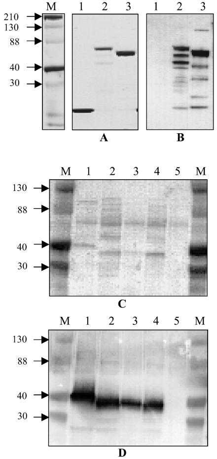
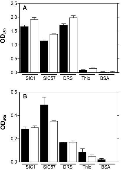
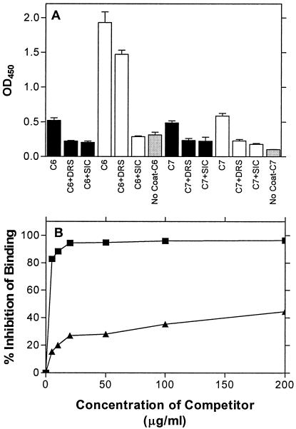
An extracellular protein of Streptococcus pyogenes, streptococcal inhibitor of complement (SIC), and its variant, called DRS (distantly related to SIC), are expressed by some S. pyogenes strains. SIC from type 1 (M1) isolates of S. pyogenes interferes with complement-mediated cell lysis, reportedly via its interaction with complement proteins. In this study we demonstrate that S. pyogenes strains carrying emm12 and emm55 (the genes for the M12 and M55 proteins, respectively) express and secrete DRS. This protein, like SIC, binds to the C6 and C7 complement proteins, and competition enzyme-linked immunosorbent assay experiments demonstrate that DRS competes with SIC for C6 and C7 binding. Similarly, SIC competes with DRS for binding to the complement proteins. Despite this, the recombinant DRS preparation showed no significant effect on complement function, as determined by lysis of sensitized sheep erythrocytes. Furthermore, the presence of DRS is not inhibitory to SIC activity.


Figures

FIG. 1.
FIG. 1.
(A and B) Recombinant DRS expressed in E. coli as thioredoxin fusion proteins. The proteins were purified as described in the text. (A) Coomassie blue-stained SDS-polyacrylamide gels. (B) Western blot reacted with a mixture (1:1) of polyclonal antibodies against SIC and DRS. Lanes 1 to 3 show His-tagged thioredoxin, SIC (BSA5)-thioredoxin fusion protein, and DRS (NS488)-thioredoxin fusion protein, respectively. The antibodies did not react with thioredoxin. (C and D) Presence of DRS in culture supernatants of DRS-expressing strains. The culture supernatants were processed as described in the text. The SDS-polyacrylamide gels were transferred to a nitrocellulose membrane (Amersham) and either stained with Ponceau S (C) or reacted with a mixture of SIC and DRS antibodies (D) as described for panel B. Lanes 1 to 5 are culture supernatants from NS844, DRV1, NS488, NS25, and NS27, respectively. NS27 is a SIC- and DRS-negative control. Channels marked M contain Kaleidoscope (Bio-Rad) markers, and approximate sizes of the proteins are shown (in kilodaltons).
FIG. 2.
FIG. 2.
Ability of DRS to bind to the C6 and C7 complement proteins. The microtiter wells were coated with proteins as indicated. The proteins were reacted with either (A) human serum or (B) complement protein C6 or C7. Binding was detected with anti-C6 (solid bars) or anti-C7 (open bars) primary antibodies.
FIG. 3.
FIG. 3.
(A) Competition of SIC binding to complement proteins by DRS and vice versa. DRS (solid bars) and SIC (open bars) were used to coat microtiter wells and reacted with (a) C6, C6 and DRS, or C6 and SIC or (b) C7, C7 and DRS, or C7 and SIC, as indicated. Each set had a no-coat control (gray bars) in which the wells were not coated with either SIC or DRS. (B) Comparison of competitive inhibition by SIC and DRS of SIC binding to the C6 protein. Wells were coated with 10 μg of SIC and reacted with C6 that had been incubated with various concentrations of SIC (▪) or DRS (▴), as indicated. In the absence of the competitors, SIC and DRS bound to the C6 protein and had an optical density at 450 nm of 0.802 and 0.787, respectively, after subtraction of the respective backgrounds (0.126 and 0.154, respectively).
FIG. 4.
FIG. 4.
Common features between SIC and DRS. Signal sequence (SS), short repeat regions (SRR), repeat regions (RR), and proline-rich regions (PR) are shown. The DRS molecule has different repeat regions (RR′) which contain short repeat region (SRR′). The similarity of the proline-rich region is indicated.

References

    1. Akesson, P., A. G. Sjoholm, and L. Bjorck. 1996. Protein SIC, a novel extracellular protein of Streptococcus pyogenes interfering with complement function. J. Biol. Chem. 271:1081-1088. - PubMed
    1. Binks, M., D. McMillan, and K. S. Sriprakash. 2003. Genomic location and variation of the gene for CRS, a complement binding protein in the M57 strains of Streptococcus pyogenes. Infect. Immun. 71:6701-6706. - PMC - PubMed
    1. Brandt, C. M., F. Allerberger, B. Spellerberg, R. Holland, R. Lutticken, and G. Haase. 2001. Characterization of consecutive Streptococcus pyogenes isolates from patients with pharyngitis and bacteriological treatment failure: special reference to prtF1 and sic/drs. J. Infect. Dis. 183:670-674. - PubMed
    1. Chatellier, S., N. Ihendyane, R. G. Kansal, F. Khambaty, H. Basma, A. Norrby-Teglund, D. E. Low, A. McGeer, and M. Kotb. 2000. Genetic relatedness and superantigen expression in group A streptococcus serotype M1 isolates from patients with severe and nonsevere invasive diseases. Infect. Immun. 68:3523-3534. - PMC - PubMed
    1. Cu, G. A., S. Mezzano, J. D. Bannan, and J. B. Zabriskie. 1998. Immunohistochemical and serological evidence for the role of streptococcal proteinase in acute post-streptococcal glomerulonephritis. Kidney Int. 54:819-826. - PubMed

Publication types

MeSH terms

LinkOut - more resources