. 2004 Jul;72(7):4159-71.
doi: 10.1128/IAI.72.7.4159-4171.2004.

Characterization of beta-glucan recognition site on C-type lectin, dectin 1

Affiliations

Characterization of beta-glucan recognition site on C-type lectin, dectin 1

Yoshiyuki Adachi et al. Infect Immun. 2004 Jul.

Abstract

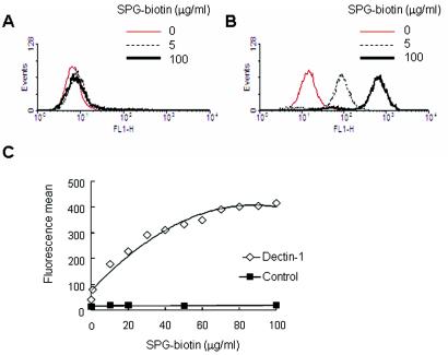
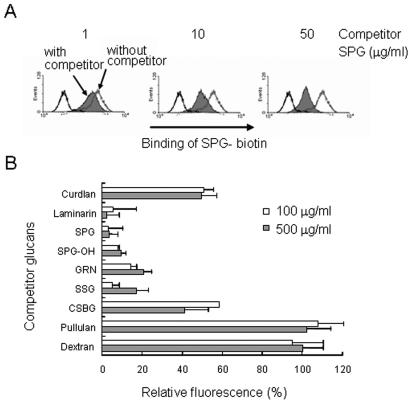
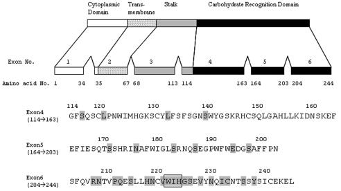
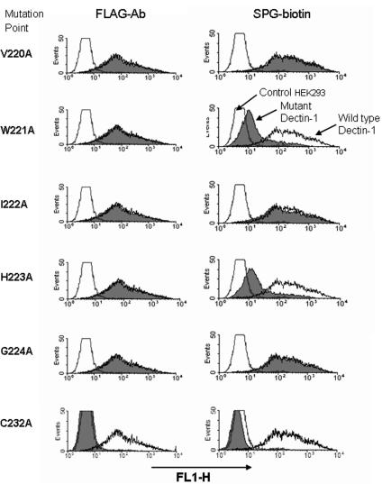
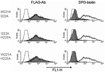
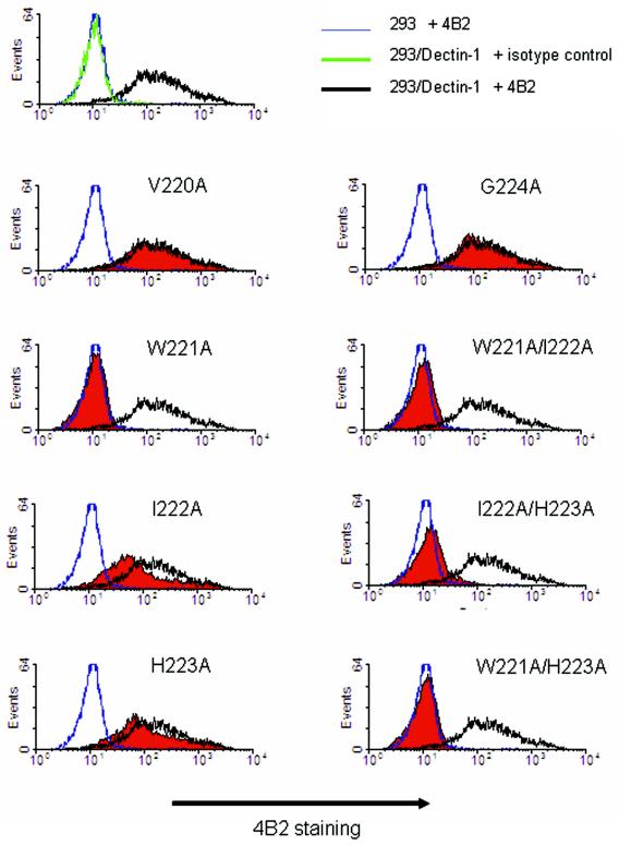
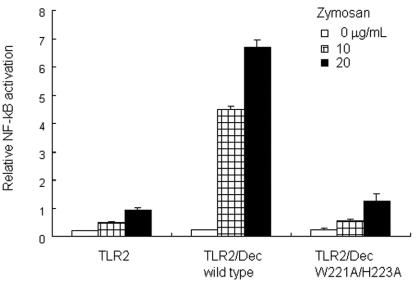
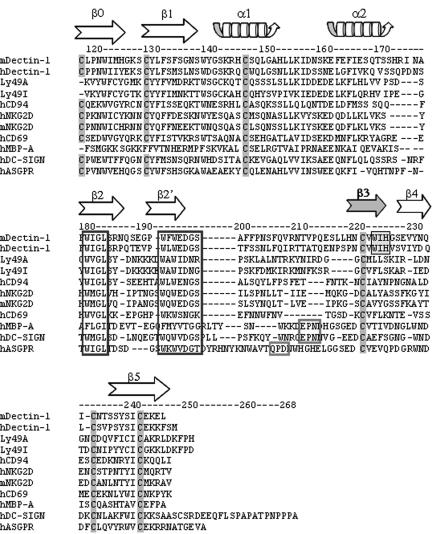
Dectin 1 is a mammalian cell surface receptor for (1-->3)-beta-d-glucans. Since (1-->3)-beta-d-glucans are commonly present on fungal cell walls, it has been suggested that dectin 1 is important for recognizing fungal invasion. In this study we tried to deduce the amino acid residues in dectin 1 responsible for beta-glucan recognition. HEK293 cells transfected with mouse dectin 1 cDNA could bind to a gel-forming (1-->3)-beta-d-glucan, schizophyllan (SPG). The binding of SPG to a dectin 1 transfectant was inhibited by pretreatment with other beta-glucans having a (1-->3)-beta-d-glucosyl linkage but not by pretreatment with alpha-glucans. Dectin 1 has a carbohydrate recognition domain (CRD) consisting of six cysteine residues that are highly conserved in C-type lectins. We prepared 32 point mutants with mutations in the CRD and analyzed their binding to SPG. Mutations at Trp(221) and His(223) resulted in decreased binding to beta-glucan. Monoclonal antibody 4B2, a dectin- 1 monoclonal antibody which had a blocking effect on the beta-glucan interaction, completely failed to bind the dectin-1 mutant W221A. A mutant with mutations in Trp(221) and His(223) did not have a collaborative effect on Toll-like receptor 2-mediated cellular activation in response to zymosan. These amino acid residues are distinct from residues in other sugar-recognizing peptide sequences of typical C-type lectins. These results suggest that the amino acid sequence W221-I222-H223 is critical for formation of a beta-glucan binding site in the CRD of dectin 1.


Figures

FIG. 1.
FIG. 1.
SPG-biotin can bind to dectin-1-transduced HEK293 cells. (A and B) Non-dectin-1A-transduced cells (A) and dectin-1-transduced cells (B) were treated with 0, 5, and 100 μg of SPG-biotin per ml. (C) Dose dependence of SPG-biotin binding to dectin-1-transduced cells or control cells, as shown by the fluorescence means from a FACS analysis. Dectin-1-transduced cells were incubated with various concentrations of SPG-biotin for 30 min on ice. After preincubation and washing, the amount of SPG-biotin bound to the cells was determined by staining with streptavidin-Alexa 488 conjugate and using a flow cytometer.
FIG. 2.
FIG. 2.
Binding of SPG-biotin to dectin-1-transduced cells can be inhibited by 1,3-β-glucans. Dectin-1A-transduced HEK293 cells were pretreated with 1, 10, and 50 μg of SPG per ml (A) or with 100 and 500 μg of various competitor glucans per ml (B) for 30 min on ice. After washing, the cells were treated with 50 μg of SPG-biotin per ml. (A) Amount of SPG binding to the cells, as shown by a histogram of flow cytometry data. (B) Amount of SPG binding quantitated by flow cytometry and expressed as the mean fluorescence relative to that of an uninhibited control (100%). The background value (0%) was obtained with nontransduced HEK293 cells treated with 50 μg of SPG-biotin per ml. The data are the averages of the means of two independent experiments, and the error bars indicate the standard errors of the means.
FIG. 3.
FIG. 3.
Schematic representation of murine dectin 1A. The residues are numbered beginning with the initial codon. The numbers 114 to 244 in the upper panel indicate positions in the CRD in the complete molecule. The sequence of amino acid residues 114 to 244, which include exons 4, 5, and 6 of murine dectin 1A, is shown in the lower panel. Mutations of single amino acid residues by Ala are indicated by shading. The boxed amino acid residues are the double-amino-acid replacement to Ala mutation (Trp/Ile, Ile/His, and Trp/His).
FIG. 4.
FIG. 4.
Effects of dectin-1 point mutations on binding to SPG-biotin. Mutant dectin 1A cDNA inserted into a FLAG-tagged expression vector was transiently transduced into HEK293 cells by using FuGene 6. The levels of expression of the FLAG tag in various dectin-1 mutants were monitored by using anti-FLAG MAb. The cells were also tested for the ability to bind SPG-biotin. The histograms show representative results for single point mutations for 29 mutants tested in this study. Reproducible results were obtained in three independent experiments. The shaded histograms show the results for mutations in dectin 1. The open histograms show the results for wild-type dectin 1 (high fluorescence) and the control vector (low fluorescence).
FIG. 5.
FIG. 5.
Double-amino-acid replacement in dectin 1 reduced binding to SPG-biotin. The levels of expression of various dectin-1 mutants were monitored by using anti-FLAG MAb. Binding of SPG-biotin to the mutants was also examined. The shaded histograms show the results for dectin-1 mutants. The open histograms show the results for wild-type dectin-1-transduced cells (high fluorescence) and control vector-transduced cells (low fluorescence).
FIG. 6.
FIG. 6.
Anti-mouse dectin-1 antibody 4B2 recognizes the glucan binding site of dectin 1. The levels of expression of various dectin-1 mutants were monitored by using anti-FLAG MAb. The cells were also assessed to determine their reactivities to dectin-1 MAb 4B2. Dectin-1-transduced cells were incubated with MAb 4B2 for 30 min on ice. After washing, anti-rat IgG-biotin for 4B2 staining and streptavidin-Alexa 488 were added and incubated for 30 min on ice. The cells were analyzed by flow cytometry after antibody staining. The shaded histograms show the results for dectin-1 mutants. The open histograms with the black, blue, and green lines show the results for wild-type dectin-1-transduced cells plus 4B2, control vector-transduced cells plus 4B2, and wild-type dectin-1-transduced cells plus isotype control antibody, respectively.
FIG. 7.
FIG. 7.
Dectin-1 mutant did not augment TLR2-mediated NF-κB activation in response to zymosan. HEK293 cells were transfected with TLR2- and dectin-1 (Dec)-expressing plasmids and an ELAM-1 promoter-conjugated luciferase reporter plasmid and were stimulated with zymosan, a yeast cell wall preparation, containing 1,3-β-glucan. NF-kB activation was monitored by the luciferase reporter assay as described in Materials and Methods. Mutation of Trp221 and His223 in the CRD of dectin 1 that resulted in replacement of both residues by alanine resulted in decreased NF-κB activation in response to zymosan.
FIG. 8.
FIG. 8.
Alignment of the dectin-1 CRD sequence with related C-type lectin CRD sequences. Highly conserved cysteine residues in CRD and β-glucan binding sites (WIH) in dectin 1 are shaded. Homologous amino acid residues involved in mannose and galactose binding and conserved amino acid sequences are enclosed in boxes. The arrows show the β-sheet strand structure of C-type lectin NK-receptors. m, mouse; h, human; MBP-A, mannose binding protein A.
FIG. 9.
FIG. 9.
Typical ultrastructure of the C-type lectin NK-receptor family. The ultrastructures of C-type lectin NK receptors CD69 (Protein Data Bank identification number, 1FM5), CD94 (Protein Data Bank identification number, 1B6E), NKG2D (Protein Data Bank identification number, 1MPU), and Ly49I (Protein Data Bank identification number, 1JA3) were visualized by using the Cn3D software, version 4.1, on the Entrez structure web server. Antiparallel β-sheets 3 and 4, which are highly conserved in other C-type lectin NK receptors, are located in the apical region of each molecule (39). Trp221 and His223 in the CRD of dectin 1 were assumed to be on β-sheet 3.

References

    1. Adachi, Y., N. Ohno, M. Ohsawa, K. Sato, S. Oikawa, and T. Yadomae. 1989. Physiochemical properties and antitumor activities of chemically modified derivatives of antitumor glucan “grifolan LE” from Grifola frondosa. Chem. Pharm. Bull. (Tokyo) 37:1838-1843. - PubMed
    1. Adachi, Y., N. Ohno, and T. Yadomae. 1993. Inhibitory effect of β-glucans on zymosan-mediated hydrogen peroxide production by murine peritoneal macrophages in vitro. Biol. Pharm. Bull. 16:462-467. - PubMed
    1. Adachi, Y., N. Ohno, and T. Yadomae. 1994. Preparation and antigen specificity of an anti-(1→3)-β-d-glucan antibody. Biol. Pharm. Bull. 17:1508-1512. - PubMed
    1. Anaissie, E. 1992. Opportunistic mycoses in the immunocompromised host: experience at a cancer center and review. Clin. Infect. Dis. 14:S43-S53. - PubMed
    1. Ariizumi, K., G. L. Shen, S. Shikano, S. Xu, R. Ritter III, T. Kumamoto, D. Edelbaum, A. Morita, P. R. Bergstresser, and A. Takashima. 2000. Identification of a novel, dendritic cell-associated molecule, dectin-1, by subtractive cDNA cloning. J. Biol. Chem. 275:20157-20167. - PubMed

Publication types

MeSH terms