

Three-dimensional virtual cholangioscopy: a reliable tool for the diagnosis of common bile duct stones
- PMID: 15213622
- PMCID: PMC1356378
- DOI: 10.1097/01.sla.0000129493.22157.b7
Three-dimensional virtual cholangioscopy: a reliable tool for the diagnosis of common bile duct stones
Abstract
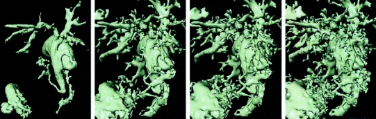
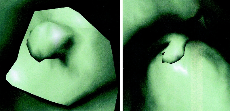
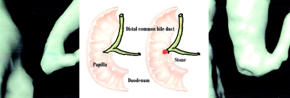
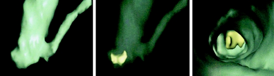
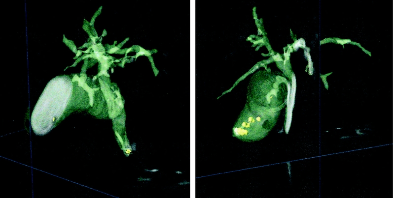
Objective: Our goal was to evaluate the clinical reliability of a new software system employing 3-dimensional (3D) virtual anatomic reconstruction and intraluminal virtual exploration for detection of choledocholithiasis and preoperative visualization of the biliary anatomy.
Summary background data: Virtual reality systems have been proposed for gastroscopy, bronchoscopy, and colonoscopy, as well as for the 3D reconstruction of liver anatomy and hepatic lesions. The impact of these systems in preoperative diagnostics has not been established due to the lack of large clinical series evaluating their reliability.
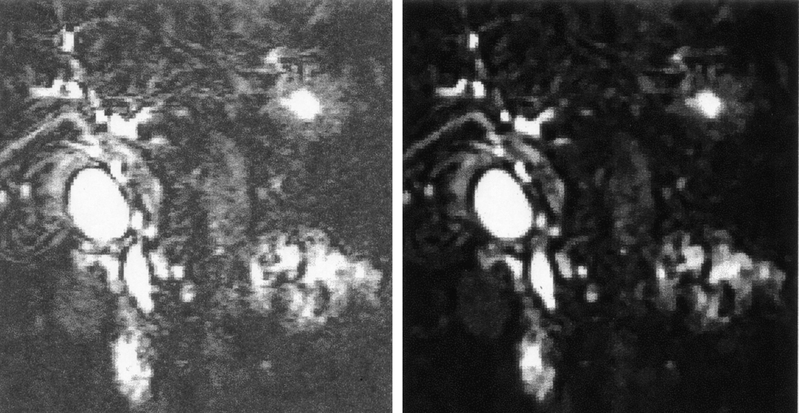
Methods: From November 2000 to July 2002, all patients presenting to our Institute with suspected choledocholithiasis were prospectively included in the study. All patients underwent conventional magnetic resonance cholangiopancreatography (MRCP) and either intraoperative cholangiogram (IOC) or endoscopic retrograde cholangiopancreatography (ERCP). The digital data from MRCP were incorporated into an original virtual reality software system to generate a 3D reconstruction. All 3D reconstructions were evaluated by a surgeon and a computer software engineer who were blind to the results of the IOC or ERCP. Sensitivity and specificity were then calculated based on the results of either the IOC or ERCP.
Results: Sixty-five patients were enrolled in the study. The average time required to reconstruct the images into navigable virtual reality was 7.5 minutes (range, 4-13.5). The 3D virtual cholangioscopy had sensitivity and specificity rates of 71% and 91%, respectively, compared with 61% and 86% of the standard MRCP.
Conclusion: : The 3D virtual cholangioscopy provides detailed preoperative reconstruction of biliary anatomy and reliable identification of choledocholithiasis with acceptable sensitivity and specificity in a clinical setting. Newer software developments may further enhance its accuracy, so that virtual cholangioscopy might challenge or replace more invasive diagnostic measures in the near future.
Figures
References
-
- Millat B, Borie F. Common bile duct stones and their complications. Rev Prat. 2000;50:2123–2129. - PubMed
-
- Hugier M. Prospective analysis of a scoring system to predict choledocholithiasis (Br J Surg 2000;87:1176–1181). Br J Surg. 2001;88:314–315. - PubMed
-
- Lincender L, Sadagic E, Vrcic D, et al. Magnetic resonance cholangiography in patients with bile duct obstruction. Radiol Oncol. 2000;34:319–324.
-
- Kyoung KT, Soo KB, Kwon HH, et al. Diagnosis of intrahepatic stones: Superiority of MR cholangiopancreatography over endoscopic retrograde cholangiopancreatography. AJR. 2002;179:429–434. - PubMed
-
- Laokpessi A, Bouillet P, Sautereau D, et al. Value of magnetic resonance cholangiography in the preoperative diagnosis of common bile duct stones. Am J Gastroenterol. 2001;96:2354–2359. - PubMed
MeSH terms
LinkOut - more resources
Full Text Sources
Medical
