Review
. 2004 Sep 15;382(Pt 3):769-81.
doi: 10.1042/BJ20040892.

Carbohydrate-binding modules: fine-tuning polysaccharide recognition

Affiliations
Review

Carbohydrate-binding modules: fine-tuning polysaccharide recognition

Alisdair B Boraston et al. Biochem J. .

Abstract

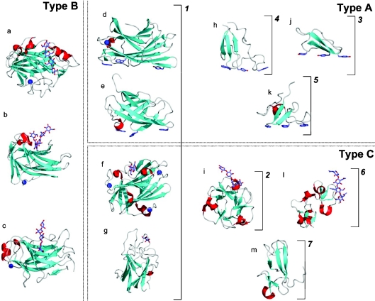
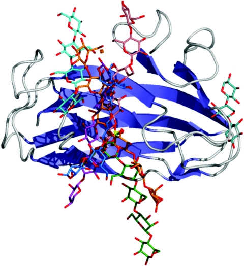
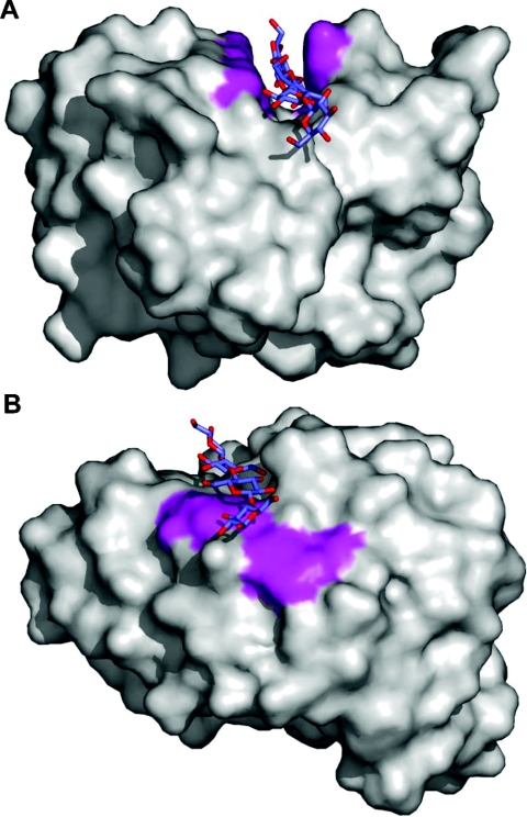
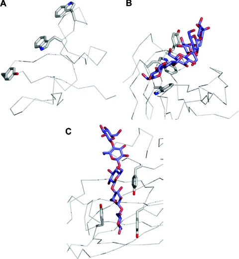
The enzymic degradation of insoluble polysaccharides is one of the most important reactions on earth. Despite this, glycoside hydrolases attack such polysaccharides relatively inefficiently as their target glycosidic bonds are often inaccessible to the active site of the appropriate enzymes. In order to overcome these problems, many of the glycoside hydrolases that utilize insoluble substrates are modular, comprising catalytic modules appended to one or more non-catalytic CBMs (carbohydrate-binding modules). CBMs promote the association of the enzyme with the substrate. In view of the central role that CBMs play in the enzymic hydrolysis of plant structural and storage polysaccharides, the ligand specificity displayed by these protein modules and the mechanism by which they recognize their target carbohydrates have received considerable attention since their discovery almost 20 years ago. In the last few years, CBM research has harnessed structural, functional and bioinformatic approaches to elucidate the molecular determinants that drive CBM-carbohydrate recognition. The present review summarizes the impact structural biology has had on our understanding of the mechanisms by which CBMs bind to their target ligands.


Figures

Figure 1
Figure 1. The big picture: CBMs, shown as ribbon structures, grouped as fold families and functional types
Dotted boxes surround examples of CBMs belonging to the functional Types A, B, and C. Brackets with numbers indicate examples of CBMs belonging to fold families 1–7. CBMs shown are as follows: (a) family 17 CBM, CcCBM17, from Clostridium cellulovorans in complex with cellotetraose (PDB code 1J84 [60]); (b) family 4 CBM, TmCBM4-2, from T. maritima in complex with laminariohexaose (PDB code 1GUI [68]); (c) family 15 CBM, CjCBM15, from Cellvibrio japonicus in complex with xylopentaose (PDB code 1GNY [67]); (d) family 3 CBM, CtCBM3, from Clostridium thermocellum (PDB code 1NBC [52]); (e) family 2 CBM, CfCBM2, from Cellulomonas fimi (PDB code 1EXG [81]); (f) family 9 CBM, TmCBM9-2, from T. maritima in complex with cellobiose (PDB code 1I82 [33]); (g) family 32 CBM, MvCBM32, from Micromonospora viridifaciens in complex with galactose (PDB code 1EUU [65]); (h) family 5 CBM, EcCBM5, from Erwinia chrysanthemi (PDB code 1AIW [82]); (i) family 13 CBM, SlCBM13, from S. lividans in complex with xylopentaose (PDB code 1MC9 [48]); (j) family 1 CBM, TrCBM1, from Trichoderma reesi (PDB code 1CBH [83]); (k) family 10 CBM, CjCBM10, from Cellvibrio japonicus (PDB code 1E8R [84]); (l) family 18 CBM from Urtica dioca in complex with chitotriose (PDB code 1EN2 [85]); (m) family 14 CBM, tachychitin, from Tachypleus tridentatus (PDB code 1DQC [86]). Bound ligands are shown as ‘liquorice’ representations, while bound metal ions are shown as a blue spheres.
Figure 2
Figure 2. Schematic diagram showing the location of binding sites in the β-sandwich Type B CBMs
This Figure was produced by overlapping the C-α carbons for all of the β-sandwich Type B CBMs for which ligand bound complexes were available. Only the ribbon structure of the family 4 CBM from the N-terminus of Cel9B from Cellulomonas fimi is shown as a representative β-sandwich Type B CBM. Oligosaccharide ligands are shown in ‘liquorice’ representation and coloured as follows: cyan, cellotetraose from CcCBM17 (PDB code 1J84 [60]); blue, laminariohexaose from TmCBM4-2 (PDB code 1GUI [68]); pink, cellopentaose from CfCBM4-2 (PDB code 1GU3 [68]); marine/aqua, xylotriose from CsCBM6-3 (PDB code 1NAE [61]); orange, cellohexaose from PeCBM29-2 (PDB code 1GWM [69]); green, mannopentaose from TmCBM27 (PDB code 1OF4 [73]); purple, xylopentaose from CjCBM15 (PDB code 1GNY [67]).
Figure 3
Figure 3. Solvent-accessible surface representations of two CBMs showing the depth of binding grooves in Type B CBMs
(A) Example of a cellopentaose molecule occupying a deep binding groove in CfCBM4-2 (PDB code 1GU3 [68]). (B) Example of a cellotetraose molecule occupying a shallow binding groove in CcCBM17 (PDB code 1J84 [60]). The surfaces created by the aromatic amino acid side chains involved in binding are shown in magenta.
Figure 4
Figure 4. The three types of binding-site ‘platforms’ formed by aromatic amino acid residues
(A) The ‘planar’ platform in the family 10 Type A CBM, CjCBM10. (B) The ‘twisted’ platform of the Type B family 29 CBM, PeCBM29-2. (C) The ‘sandwich’ platform of the Type B family 4 CBM, CfCBM4-2. The C-α backbone is shown as grey cylinders, the aromatic amino acid side chains forming the binding sites are shown in grey ‘liquorice’ representations, and the bound oligosaccharides are shown in blue ‘liquorice’ representation.
Figure 5
Figure 5. Binding-site topography and oligosaccharide recognition
Surface representations of the NMR structures of wild-type CfCBM2b-1 (A) and the same protein with an Arg262→Gly mutation (B). The arrow indicates the tryptophan residue that changes conformation due to the mutation. The binding site of CfCBM4-1 extend in a straight path across the face of one of the β-sheets of this CBM creating a linear binding site appropriate for binding cello-oligosaccharides (C). In TmCBM4-2, which has significant sequence identity with CfCBM4-1, two loops are extended (D); one to block an end of the binding site (shown by the arrow labelled 1) and another to accommodate the curvature of the laminarioligosaccharide (shown by the arrow labelled 2). Solvent-accessible surfaces are shown in transparent grey with surfaces created by the aromatic amino acid side chains involved in binding shown in magenta. C-α traces are shown in blue.
Figure 6
Figure 6. The role of calcium in xylan recognition by the family 36 CBM, PpCBM36, from Pa. polymyxa xylanase 43
The calcium atom bound in the binding site of PpCBM36 is shown as a blue sphere. The amino acid residues involved in co-ordinating the calcium and binding the sugar are shown in grey ‘liquorice’ representation, the bound xylo-oligosaccharide is shown in blue ‘liquorice’ representation and the C-α backbone is represented as grey cylinders.
Figure 7
Figure 7. The accommodation of galactomannan by a family 27 CBM, TmCBM27
The overall structure surrounding the binding site of TmCBM27 from T. maritima is shown as a ribbon diagram. The three tryptophan side chains comprising the carbohydrate-binding site are shown in grey ‘liquorice’ representation and are numbered. The ligand, G2M5, a fragment of galactomannan, is shown in ‘liquorice’ representation with mannose residues in blue and galactose residues in yellow. The uppermost galactose residue in the Figure is clearly bent back over the top of the mannose backbone by Trp28, while the other galactose residue extends comfortably away from the mannose backbone.
Figure 8
Figure 8. Multivalent CBMs
The family 13 CBM from S. lividans xylanase 10A, SlCBM13, is shown in complex with three lactose molecules occupying its three binding sites (A), while the family 6 CBM from Cellvibrio mixtus endoglucanase 5A, CmCBM6, is shown in complex with two cellobiose molecules occupying its two binding sites (B). Both are shown as ribbon diagrams with relevant aromatic amino acid side chains in the binding sites shown in grey ‘liquorice’ representation. The three β-trefoil subdomains of SlCBM13 are labelled as α, β and γ, and the two binding sites of CmCBM6 are labelled according to the cleft in which they reside.

References

    1. Van Tilbeurgh H., Tomme P., Claeyssens M., Bhikhabhai R., Pettersson G. Limited proteolysis of the cellobiohydrolase I from Trichoderma reesei. FEBS Lett. 1986;204:223–227.
    1. Tomme P., Van Tilbeurgh H., Pettersson G., Van Damme J., Vandekerckhove J., Knowles J., Teeri T., Claeyssens M. Studies of the cellulolytic system of Trichoderma reesei QM 9414: analysis of domain function in two cellobiohydrolases by limited proteolysis. Eur. J. Biochem. 1988;170:575–581. - PubMed
    1. Gilkes N. R., Warren R. A. J., Miller R. C., Jr, Kilburn D. G. Precise excision of the cellulose binding domains from two Cellulomonas fimi cellulases by a homologous protease and the effect on catalysis. J. Biol. Chem. 1988;263:10401–10407. - PubMed
    1. Boraston A. B., McLean B. W., Kormos J. M., Alam M., Gilkes N. R., Haynes C. A., Tomme P., Kilburn D. G., Warren R. A. J. Carbohydrate-binding modules: diversity of structure and function. In: Gilbert H. J., Davies G. J., Henrissat B., Svensson B., editors. Recent Advances in Carbohydrate Bioengineering. Cambridge: Royal Society of Chemistry; 1999. pp. 202–211.
    1. Coutinho P. M., Henrissat B. Carbohydrate-active enzymes: an integrated database approach. In: Gilbert H. J., Davies G. J., Henrissat B., Svensson B., editors. Recent Advances in Carbohydrate Bioengineering. Cambridge: Royal Society of Chemistry; 1999. pp. 3–12.