Comparative Study
. 2004 Jul;165(1):35-52.
doi: 10.1016/S0002-9440(10)63273-7.

Inhibition of vascular endothelial growth factor (VEGF) signaling in cancer causes loss of endothelial fenestrations, regression of tumor vessels, and appearance of basement membrane ghosts

Affiliations
Comparative Study

Inhibition of vascular endothelial growth factor (VEGF) signaling in cancer causes loss of endothelial fenestrations, regression of tumor vessels, and appearance of basement membrane ghosts

Tetsuichiro Inai et al. Am J Pathol. 2004 Jul.

Abstract

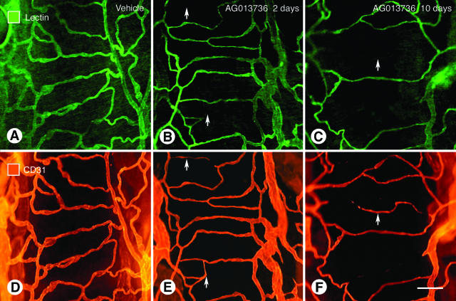
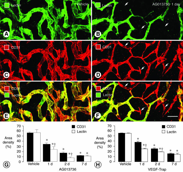
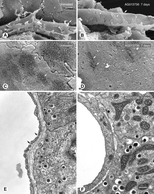
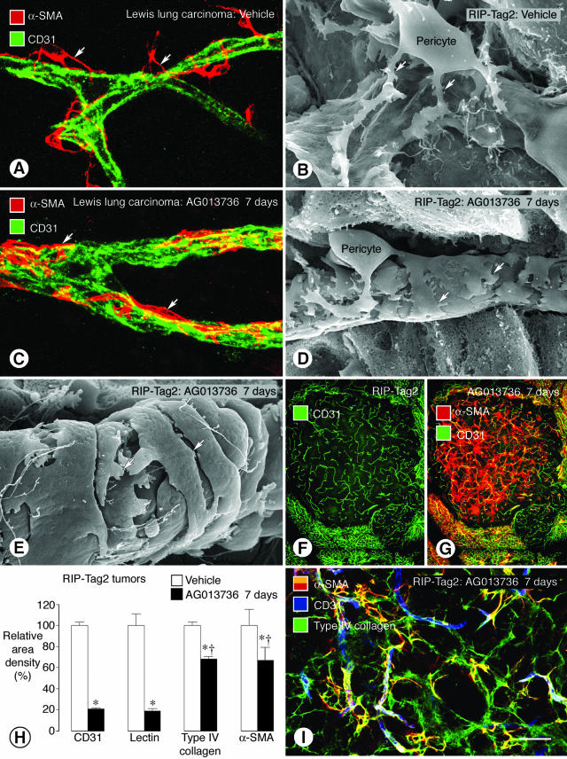
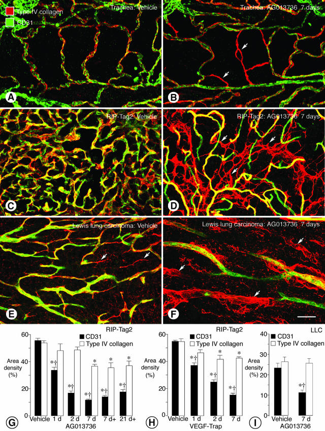
Angiogenesis inhibitors are receiving increased attention as cancer therapeutics, but little is known of the cellular effects of these inhibitors on tumor vessels. We sought to determine whether two agents, AG013736 and VEGF-Trap, that inhibit vascular endothelial growth factor (VEGF) signaling, merely stop angiogenesis or cause regression of existing tumor vessels. Here, we report that treatment with these inhibitors caused robust and early changes in endothelial cells, pericytes, and basement membrane of vessels in spontaneous islet-cell tumors of RIP-Tag2 transgenic mice and in subcutaneously implanted Lewis lung carcinomas. Strikingly, within 24 hours, endothelial fenestrations in RIP-Tag2 tumors disappeared, vascular sprouting was suppressed, and patency and blood flow ceased in some vessels. By 7 days, vascular density decreased more than 70%, and VEGFR-2 and VEGFR-3 expression was reduced in surviving endothelial cells. Vessels in Lewis lung tumors, which lacked endothelial fenestrations, showed less regression. In both tumors, pericytes did not degenerate to the same extent as endothelial cells, and those on surviving tumor vessels acquired a more normal phenotype. Vascular basement membrane persisted after endothelial cells degenerated, providing a ghost-like record of pretreatment vessel number and location and a potential scaffold for vessel regrowth. The potent anti-vascular action observed is evidence that VEGF signaling inhibitors do more than stop angiogenesis. Early loss of endothelial fenestrations in RIP-Tag2 tumors is a clue that vessel phenotype may be predictive of exceptional sensitivity to these inhibitors.


Figures

Figure 1
Figure 1
Loss of vessel patency in simple vascular network of trachea after inhibition of VEGF signaling. Fluorescence micrographs showing vasculature of mouse tracheas stained by intravenous injection of lectin (A–C) and same vessels stained for CD31 immunoreactivity (D and E). Three conditions are compared: A and D, normal state; B and E, after 2 days of treatment with AG013736; and C and F, after 10 days of treatment with AG013736. After 2 days of treatment the lectin stains fewer vessels than CD31 because vessel patency is lost and blood flow stops before the CD31 disappears as endothelial cells degenerate. At 10 days, fewer vessels are evident by either method. Arrows mark regions where the lectin staining and CD31 immunoreactivity do not match. Scale bar, 30 μm.
Figure 2
Figure 2
Reduction in tumor vessel patency and blood flow after inhibition of VEGF signaling. Confocal microscopic images of tumor vessels in RIP-Tag2 mice treated with vehicle (A, C, E) or AG013736 (B, D, F) for 1 day and injected with lectin before fixation by vascular perfusion. Lectin staining (A, green) of blood vessels in vehicle-treated tumor closely matches CD31 immunoreactivity (C, red). However, after treatment with AG013736, multiple vessels (arrows) lack lectin staining (B, green) but have CD31 immunoreactivity (D, red), indicating loss of vessel patency. Vessels without flow appear red in merged images (E, F). Bar graphs show area density (%) of lectin staining and CD31 immunoreactivity in vessels of RIP-Tag2 tumors after treatment for 1, 2, or 7 days with AG013736 (G) or VEGF-Trap (H) or their respective vehicle. After 1 or 2 days of treatment by either agent, the area density of lectin staining is significantly less than the amount of CD31 immunoreactivity. By 7 days, this discrepancy no longer exists, suggesting that the surviving vessels have blood flow. *, Different from vehicle (P < 0.05). †, Different from CD31 (P < 0.05). Scale bar, 25 μm.
Figure 3
Figure 3
Reduction in endothelial sprouts and fenestrations in RIP-Tag2 tumors after inhibition of VEGF signaling. A and B: SEM of outer surface of tumor vessels showing endothelial sprouts (arrowheads) without treatment (A) but not after treatment with AG013736 for 7 days (B). Processes of pericytes are also visible (arrows), those without treatment being more loosely associated with the endothelium than after treatment. C and D: Higher magnification SEM showing much more abundant endothelial fenestrations (arrows) on abluminal surface of untreated tumor vessel (C) than after treatment (D, AG013736, 7 days). E and F: TEM showing abundance of endothelial fenestrations (arrows) in untreated tumor vessel (E) compared to none after treatment (F, AG013736, 1 day). In sections of tumor vessels examined by TEM, 63% and 83% had no fenestrations in the 1- and 7-day AG013736 groups, respectively, but only 7% had none without treatment. Scale bar: 5 μm (A, B); 1 μm (C, D); 0.5 μm (E, F).
Figure 4
Figure 4
Reduction in brightness of receptor immunofluorescence and decrease in vessel number after inhibition of VEGF signaling. Fluorescence micrographs comparing high VEGFR-2 immunoreactivity and high vessel density in vehicle-treated RIP-Tag2 tumor (A) with low VEGFR-2 immunoreactivity and low vessel density after AG013736 treatment for 7 days (B). Reduced brightness of VEGFR-2 immunoreactivity is illustrated by lower peaks in the surface plot of fluorescence intensity (C). Reduced vessel number is indicated by fewer peaks (C). By contrast, after staining for CD105 immunoreactivity, vessel brightness is not reduced by the treatment, but vessel number is clearly reduced (D–F). Consistent with molecule-specific changes in expression levels indicated by brightness of fluorescence, intensity measurements show significant reductions in VEGFR-2, VEGFR-3, and α5-integrin immunoreactivities of RIP-Tag2 tumor vessels after treatment with AG013736 for 7 days but little or no reduction in CD105, CD31, or PDGFR-β (G, values for vehicle normalized to 100%). VEGF-Trap for 7 days also reduced brightness of VEGFR-2 immunofluorescence in RIP-Tag2 tumor vessels, but the change was smaller (H, values for vehicle normalized to 100%). AG013736 had a smaller effect (−16%) on brightness of VEGFR-2 immunofluorescence in LLCs than found in RIP-Tag2 tumors, both in fluorescence micrographs (I, J) and surface plot (K). *, Different from corresponding vehicle (P < 0.05). †, Different from VEGF-Trap (P < 0.05). Scale bar, 80 μm.
Figure 5
Figure 5
Tightening of pericytes on tumor vessels after inhibition of VEGF signaling. Confocal microscopic and SEM images show changes in pericyte-endothelial cell relationships (arrows) in tumor vessels. In vessels of LLC, α-SMA immunoreactive pericytes (A, red) are loosely attached to CD31-positive endothelial cells (green). A similar loose association of pericytes to endothelial cells is also evident by SEM in RIP-Tag2 tumor vessels (B). By comparison, after treatment with AG013736 for 7 days, α-SMA-positive pericytes are tighter on endothelial cells imaged by immunofluorescence (C) or SEM (D). Pericytes on some treated vessels acquire a smooth muscle-like phenotype (E). RIP-Tag2 tumor vessels still present after AG013736 for 7 days (F) are accompanied by a disproportionately large number of α-SMA-positive cells, many of which are not associated with blood vessels (G). After treatment with AG013736, reductions in α-SMA and type IV collagen immunoreactivities are about the same but are much less than corresponding reductions in CD31 and lectin (H, values for vehicle normalized to 100%). After the ∼80% reduction in RIP-Tag2 tumor vessels treated with AG013736 for 7 days, most α-SMA-positive cells are still covered by type IV collagen basement membrane (I). *, Different from corresponding vehicle (P < 0.05). †, Different from CD31. Scale bar: 20 μm (A, C); 5 μm (B, D); 500 μm (F, G); 50 μm (I).
Figure 6
Figure 6
Persistence of vascular basement membrane after endothelial cells degenerate. Confocal micrographs show co-localization of CD31 immunoreactivity (green) and type IV collagen immunoreactivity (red) on simple vasculature of normal adult mouse trachea (A). After treatment with AG013736 for 7 days, sleeves of type IV collagen immunoreactivity devoid of CD31 immunoreactive endothelial cells are present among blood vessels of the tracheal mucosa (B, arrows). Similarly, in vehicle-treated RIP-Tag2 tumors, CD31 and type IV collagen immunoreactivities co-localize on most vessels (C), but after treatment with AG013736 for 7 days, empty sleeves of type IV collagen immunoreactivity are abundant (D, arrows). In LLC, some type IV collagen sleeves are present without treatment (E, arrows) but are much more numerous after treatment (F, arrows). Measurements show that area densities of CD31 and type IV collagen immunoreactivities were about the same in vehicle-treated RIP-Tag2 tumors, but after treatment with AG013736 for 1 to 21 days (d, twice daily; d+, once daily), CD31 immunoreactivity decreased much more than type IV collagen (G). This difference was detectable even at 1 day. A similar discrepancy was found after treatment with VEGF-Trap (H). In LLC, type IV collagen immunoreactivity remained essentially unchanged after treatment with AG013736 for 7 days despite the 52% reduction in CD31 staining (I). *, Different from corresponding vehicle (P < 0.05). †, Different from type IV collagen. Scale bar: 30 μm (A, B); 50 μm (C–F).

References

    1. Hlatky L, Hahnfeldt P, Folkman J. Clinical application of antiangiogenic therapy: microvessel density, what it does and doesn’t tell us. J Natl Cancer Inst. 2002;94:883–893. - PubMed
    1. Herbst RS, Mullani NA, Davis DW, Hess KR, McConkey DJ, Charnsangavej C, O’Reilly MS, Kim HW, Baker C, Roach J, Ellis LM, Rashid A, Pluda J, Bucana C, Madden TL, Tran HT, Abbruzzese JL. Development of biologic markers of response and assessment of antiangiogenic activity in a clinical trial of human recombinant endostatin. J Clin Oncol. 2002;20:3804–3814. - PubMed
    1. Ellis LM. Antiangiogenic therapy: more promise and, yet again, more questions. J Clin Oncol. 2003;21:3897–3899. - PubMed
    1. McCarthy M. Antiangiogenesis drug promising for metastatic colorectal cancer. Lancet. 2003;361:1959. - PubMed
    1. Yang JC, Haworth L, Sherry RM, Hwu P, Schwartzentruber DJ, Topalian SL, Steinberg SM, Chen HX, Rosenberg SA. A randomized trial of bevacizumab, an anti-vascular endothelial growth factor antibody, for metastatic renal cancer. N Engl J Med. 2003;349:427–434. - PMC - PubMed

Publication types

MeSH terms