. 2004 Jul;165(1):115-26.
doi: 10.1016/s0002-9440(10)63280-4.

Nuclear factor-kappaB regulates inflammatory cell apoptosis and phagocytosis in rat carrageenin-sponge implant model

Affiliations

Nuclear factor-kappaB regulates inflammatory cell apoptosis and phagocytosis in rat carrageenin-sponge implant model

Maria Chiara Maiuri et al. Am J Pathol. 2004 Jul.

Abstract

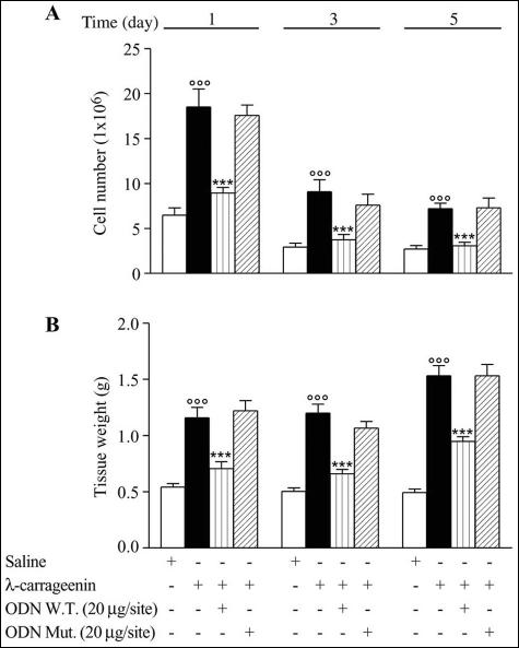
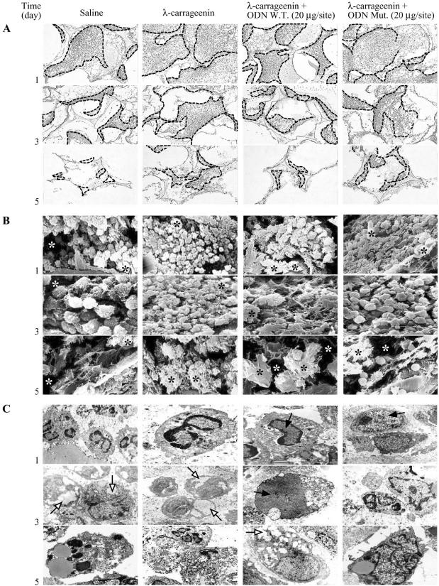
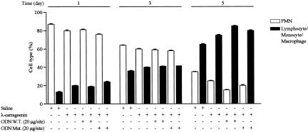
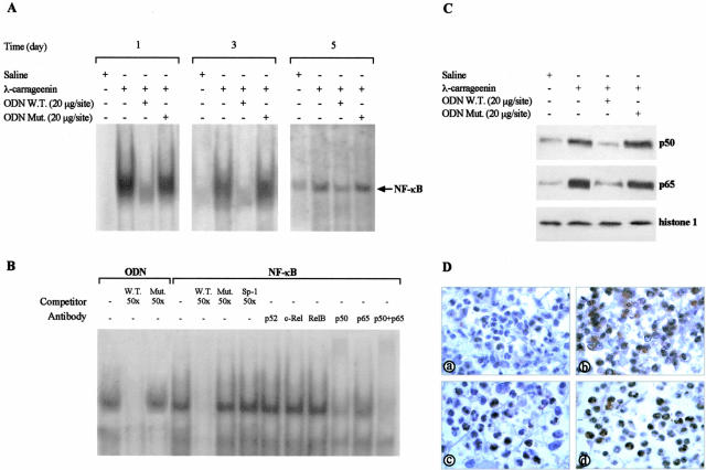
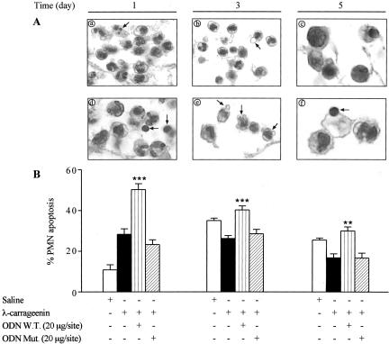
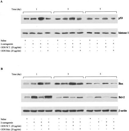
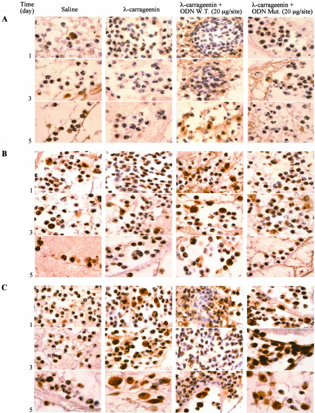
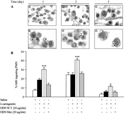
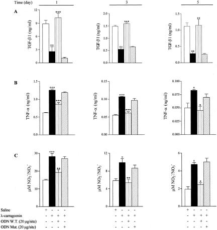
In the present study we investigated whether apoptosis and phagocytosis are regulated by nuclear factor (NF)-kappaB in a model of chronic inflammation. The subcutaneous implant of lambda-carrageenin-soaked sponges elicited an inflammatory response, characterized by a time-related increase of leukocyte infiltration into the sponge and tissue formation, which was inhibited by simultaneous injection of wild-type oligodeoxynucleotide decoy to NF-kappaB. Molecular and morphological analysis performed on infiltrated cells demonstrated: 1) an inhibition of NF-kappaB/DNA binding activity; 2) an increase of polymorphonuclear leukocyte apoptosis correlated either to an increase of p53 or Bax and decrease of Bcl-2 protein expression; and 3) an increase of phagocytosis of apoptotic polymorphonuclear leukocytes by macrophages associated with an increase of transforming growth factor-beta1 and decrease of tumor necrosis factor-alpha as well as nitrite/nitrate production. Our results, showing that blockade of NF-kappaB by oligodeoxynucleotide decoy increases inflammatory cell apoptosis and phagocytosis, may contribute to lead to new insights into the mechanisms governing the inflammatory process.


Figures

Figure 1
Figure 1
Leukocyte infiltration (A) and granulomatous tissue formation (B). Data are expressed as mean ± SEM of 15 to 20 sponges from 9 to 10 rats. °°°, P < 0.0001 versus saline; ***, P < 0.0001 versus λ-carrageenin alone.
Figure 2
Figure 2
Morphological summary depicting cell migration, tridimensional organization, and activation status in the sponge after treatments. A: Light microscopy: relationship between spaces invaded by cells (outlined areas) and exudate as well as debris. B: Scanning electron microscopy: presence of cells organized even in clusters with debris (black asterisk) and exudate (white asterisk) areas. C: Transmission electron microscopy: ultrastructural features of cell populations with cytosol suggestive of phagocytosis as well as autophagy (open arrows) and nuclei involution (filled arrows) Fields are representative of three experiments. Original magnifications: ×100 (A); ×2400 (B); ×6400 to 8900 (C).
Figure 3
Figure 3
Leukocyte population infiltrated into the sponge. Data are expressed as mean ± SEM of three sponges from three rats. Cell counting was performed as reported in Materials and Methods.
Figure 4
Figure 4
A: Representative electrophoretic mobility shift assay showing NF-κB/DNA binding activity in nuclear extract from inflammatory cells. Data are from a single experiment and are representative of six separate experiments. B: Characterization of NF-κB/DNA complex was performed on λ-carrageenin-treated cells harvested on 1 day after implant. In competition reaction nuclear extracts were incubated with radiolabeled W.T. ODN probe in the absence or presence of identical but unlabeled oligonucleotide (W.T., 50×) or mutated nonfunctional ODN probe (Mut, 50×). In addition, nuclear extracts were incubated with radiolabeled NF-κB probe in absence or presence of: identical but unlabeled oligonucleotide (W.T., 50×), mutated nonfunctional NF-κB probe (Mut, 50×) or unlabeled oligonucleotide containing the consensus sequence for Sp-1 (Sp-1, 50×). In supershift experiments nuclear extracts were incubated with antibodies against p52, c-Rel, RelB, p50, and p65 15 minutes before incubation with radiolabeled NF-κB probe. Data are from a single experiment and are representative of six separate experiments. C: Representative Western blot of p50 and p65 nuclear level from cells collected on 1 day after implant. Histone 1 expression is shown as control. Data are from a single experiment and are representative of three separate experiments. D: Immunohistochemical analysis of p65 protein expression in cells from saline (a), λ-carrageenin (b), W.T. ODN (c), and Mut ODN (d) decoy-treated sponges harvested on 1 day after implant. Fields are representative of three experiments. Original magnifications, ×320 (D).
Figure 5
Figure 5
A: Morphological features of apoptosis (arrows) in PMNs from λ-carrageenin (ac) and W.T. ODN decoy-treated sponges (df). Fields are representative of three experiments. B: PMN apoptosis was evaluated by FACS using annexin V staining. Data are expressed as mean ± SEM of three to five sponges from three to five rats. **, P < 0.001; ***, P < 0.0001 versus λ-carrageenin alone. Original magnifications, ×320 (A).
Figure 6
Figure 6
Representative Western blot of p53 (A), Bax, and Bcl-2 (B) protein expression in nuclear and cytosolic extracts from inflammatory cells. Histone 1 and β-actin expression are shown as control, respectively. Data are from a single experiment and are representative of three to four separate experiments.
Figure 7
Figure 7
Immunohistochemical summary of p53 (A), Bax (B), and Bcl-2 (C) protein expression in inflammatory cells after treatments. Fields are representative of three experiments. Original magnifications, ×320.
Figure 8
Figure 8
A: Morphological appearance of MΦ phagocytosis (arrows) from λ-carrageenin (a–c) and W.T. ODN decoy-treated sponges (d–f). Fields are representative of three experiments. B: Phagocytosis was evaluated as phagocytic percent. Data are expressed as percentage of mean ± SEM of MΦ containing at least one ingested apoptotic PMN (n = 3 sponges from three rats). *, P < 0.05; ***, P < 0.0001 versus λ-carrageenin alone. Original magnifications, ×320 (A).
Figure 9
Figure 9
Effect of W.T. ODN decoy on TGF-β1 (A), TNF-α (B), and NOx (C) production by inflammatory exudate. Data are expressed as mean ± SEM of three experiments in triplicate. °, P < 0.05; °°, P < 0.001; °°°, P < 0.0001 versus saline; *, P < 0.05; **, P < 0.001; ***P < 0.0001 versus λ-carrageenin alone.

References

    1. Savill J. Apoptosis in resolution of inflammation. J Leukoc Biol. 1997;61:375–380. - PubMed
    1. Kobayashi SD, Voyich JM, Buhl CL, Stahl RM, DeLeo FR. Global changes in gene expression by human polymorphonuclear leukocytes during receptor-mediated phagocytosis: cell fate is regulated at the level of gene expression. Proc Natl Acad Sci USA. 2002;99:6901–6906. - PMC - PubMed
    1. Kobayashi SD, Voyich JM, Somerville JA, Braughton KR, Malech HL, Musser JM, DeLeo FR. An apoptosis-differentiation program in human polymorphonuclear leukocytes facilitates resolution of inflammation. J Leukoc Biol. 2003;73:315–322. - PubMed
    1. Savill J, Dransfield I, Gregory C, Haslett C. A blast from the past: clearance of apoptotic cells regulates immune responses. Nat Rev. 2002;2:965–975. - PubMed
    1. Savill J, Wyllie AH, Henson JE, Walport MJ, Henson PM, Haslett C. Macrophage phagocytosis of aging neutrophils in inflammation. Programmed cell death in the neutrophil leads to its recognition by macrophages. J Clin Invest. 1989;83:865–875. - PMC - PubMed

Publication types

MeSH terms