Comparative Study
. 2004 Jul;165(1):247-58.
doi: 10.1016/s0002-9440(10)63293-2.

Regulation of macrophage foam cell formation by alphaVbeta3 integrin: potential role in human atherosclerosis

Affiliations
Comparative Study

Regulation of macrophage foam cell formation by alphaVbeta3 integrin: potential role in human atherosclerosis

Alexander S Antonov et al. Am J Pathol. 2004 Jul.

Abstract

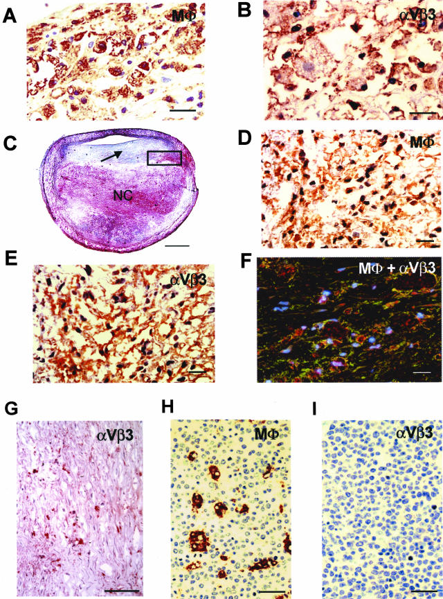
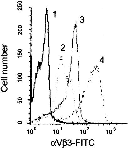
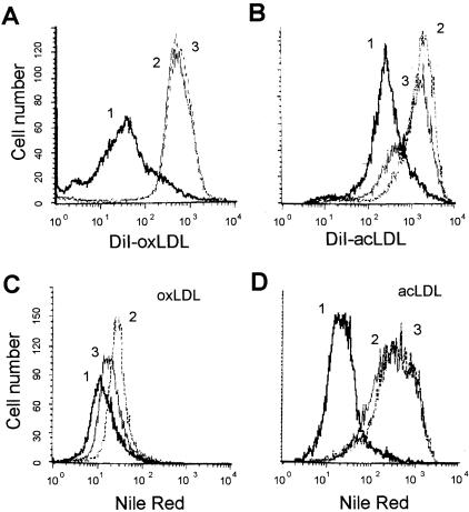
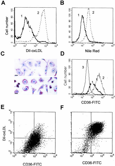
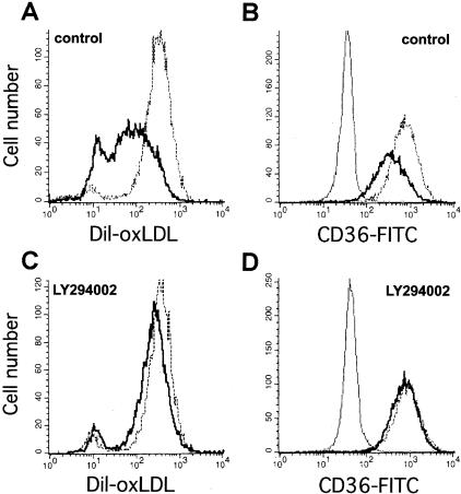
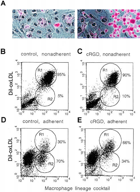
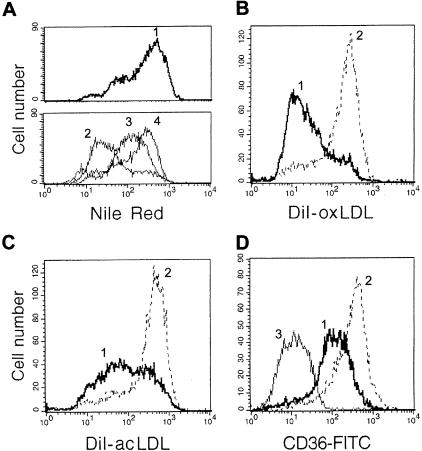
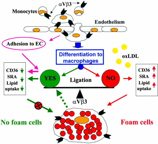
The accumulation of macrophage foam cells in atherosclerotic lesions is associated with both initiation and progression of this disease. Scavenger receptors CD36 and SRA are the primary receptors responsible for conversion of macrophages into foam cells. Integrin alphaVbeta3 plays a role in the differentiation of several cell types, but its involvement in the transition of macrophages into foam cells and the potential role of this receptor in atherosclerosis have not been examined. Using an in vitro model of single surface receptor activation by binding with an immobilized monoclonal antibody specific to alphaVbeta3 integrin we show that ligation of alphaVbeta3 integrin prevents differentiation of blood monocytes and macrophages into the foam cell phenotype via coordinate down-regulation of CD36 and SRA. This effect of alphaVbeta3 integrin ligation can be reproduced by contact with endothelial cells, whereas the inhibition of alphaVbeta3 receptor ligation restores the uptake of oxidized low-density lipoprotein. Moreover, we found that alphaVbeta3 integrin is readily detected in situ on macrophages in early and advanced atherosclerotic lesions and that in vitro exposure to oxidized low-density lipoprotein up-regulates alphaVbeta3 integrin expression. We hypothesize that alphaVbeta3 integrin regulates macrophage functional maturation into foam cells in a persistent manner, and therefore, by targeting alphaVbeta3 receptor it could potentially be possible to regulate progression of atherosclerosis in humans.


Figures

Figure 1
Figure 1
Expression of αVβ3 integrin on macrophages in a human atherosclerotic lesion. A: Accumulation of HAM-56-positive macrophages in early lipid lesion. B: Macrophages in the same lesion show strong positive staining for αVβ3 integrin expression (brownish-red reaction product). C: Low-power view of a human atherosclerotic plaque (fibroatheroma) from mid left anterior descending coronary artery. There is a large necrotic core (NC) and relatively thick fibrous cap (arrow). D: High-power view of the black box represented in C demonstrating numerous CD68-positive macrophages in the perimeter of the necrotic core. E: Positive staining for αVβ3 integrin expression on macrophages in a similar area as in D. F: Image from confocal microscopy showing co-localization of macrophages (red) and αVβ3 integrin (green) in the perimeter of the necrotic core. Regions of overlap appear yellow. The nuclei (blue) are stained with 4,6-diamidino-2-phenylindole. G: Immunostaining for αVβ3 integrin expression on SMCs within the fibrous cap of this lesion shows a relatively low number of αVβ3-positive SMCs. H: HAM-56-positive macrophages in tonsil. I: Absence of staining for αVβ3 integrin in macrophage in tonsil. Scale bars: 20 μm (A, B, D–F), 1 mm (C), 100 μm (G), 29 μm (H and I).
Figure 2
Figure 2
M-CSF and oxLDL synergistically up-regulate αVβ3 integrin surface expression on MDMs. Differentiation of freshly isolated blood monocytes in the presence of M-CSF for 6 days results in increases in αVβ3 expression on MDMs (compare lines 2 and 3). Treatment of these MDMs with oxLDL (20 μg/ml) for 24 hours significantly up-regulates αVβ3 integrin expression (line 4). Monocytes and harvested MDMs were stained for αVβ3 integrin surface expression with FITC-labeled anti-αVβ3 integrin mAb or FITC-labeled isotype-matched control IgG1 (line 1) and analyzed by flow cytometry (refer to Materials and Methods).
Figure 3
Figure 3
Differentiation of blood monocytes on immobilized anti-αVβ3 integrin mAb results in down-regulation of DiI-oxLDL and DiI-acLDL uptake and resultant lipid accumulation induced by modified LDL. Blood monocytes were seeded directly on immobilized anti-αVβ3 integrin mAb (thick line), anti-αVβ5 integrin mAb (thin line), or control isotype-matched IgG1 (dotted line) and were cultured for 7 days. On day 7 MDMs were incubated with 5 μg/ml of DiI-oxLDL (A) or 5 μg/ml of DiI-acLDL (B) for 3 hours. To induce lipid accumulation in cholesterol storage vacuoles, MDMs were exposed to 50 μg/ml of oxLDL (C) or 100 μg/ml of acLDL (D) for 48 hours (from day 5 to day 7). At the end of the treatment cells were harvested, stained for intracellular lipids with Nile Red (C and D only), and analyzed by flow cytometry.
Figure 4
Figure 4
Ligation of αVβ3 integrin on differentiated MDMs decreases DiI-oxLDL uptake and prevents oxLDL-induced foam cell formation via a mechanism requiring down-regulation of CD36 expression. MDMs (5 to 9 days of culture in vitro) were harvested and reseeded on immobilized antibodies to αVβ3 integrin (thick line) or control isotype-matched IgG1 (dotted line), and analyzed by flow cytometry 24 hours after reseeding. A: DiI-oxLDL uptake by MDMs after 3 hours of incubation with 5 μg/ml of labeled oxLDL. B: Lipid accumulation in cholesterol storage vacuoles induced by incubation with 50 μg/ml of oxLDL for 48 hours revealed by Nile Red staining. C: Reduction of foam cell formation in vitro induced by incubation with oxLDL (50 μg/ml) for 48 hours of MDMs reseeded on immobilized anti-αVβ3 mAb. Top: MDMs reseeded on immobilized anti-αVβ3 mAb. Bottom: MDM reseeded on immobilized control IgG1. Oil Red O lipid staining (red), hematoxylin nuclear counterstain (blue). D: Down-regulation of CD36 surface expression on MDMs induced by αVβ3 ligation revealed by staining with FITC-labeled mAb to CD36 and after analysis by flow cytometry (thin line shows isotype-matched control). E and F: CD36 surface expression (x axis) and DiI-oxLDL uptake (y axis) by MDMs reseeded on αVβ3 integrin antibodies (E) or control IgG1 (F). Scale bars, 20 μm.
Figure 5
Figure 5
Inhibition of PI3-kinase/Akt signaling pathway abolishes the down-regulation of DiI-oxLDL uptake and CD36 expression induced by αVβ3 integrin ligation on MDMs. MDMs were differentiated in vitro for 6 days, harvested, incubated in suspension with LY294002 (10 μmol/L) or control vehicle (dimethyl sulfoxide) for 20 minutes, and reseeded on immobilized antibodies to αVβ3 integrin (thick line) or control IgG1 (dotted line) for 6 hours in the presence of inhibitor. MDMs were incubated with DiI-oxLDL (5 μg/ml) for 3 more hours, then DiI-oxLDL uptake was analyzed by flow cytometry. For CD36 detection, MDMs reseeded on anti-αVβ3 or control mAbs were analyzed by flow cytometry using FITC-labeled mAb to CD36 or FITC-conjugated isotype control antibody (thin line). A: DiI-oxLDL uptake. B: CD36 expression by MDMs treated with vehicle control. C and D: Treatment of MDMs with LY294002 prevents effects of αVβ3 integrin ligation on DiI-oxLDL uptake (C) and CD36 expression (D).
Figure 6
Figure 6
Adhesion of MDMs to ECs prevents foam cell formation and down-regulates DiI-oxLDL uptake via mechanisms requiring αVβ3 integrin. A: Monocytes were co-cultured with ECs for 5 days, then EC monolayers were partially denuded with a rubber scraper to allow floating MDMs from co-culture to adhere to the plastic in the denuded area. Foam cell formation was induced by incubation of co-cultures with oxLDL (50 μg/ml) for 48 hours. MDMs adherent to ECs (left, arrows) show low lipid accumulation compared to those adherent to denuded area (right). Oil Red O lipid staining (red), hematoxylin counterstain (blue). B–E: Effect of the αVβ3 inhibitor cRGD on DiI-oxLDL uptake by MDMs adherent or nonadherent to ECs. Monocytes were co-cultured with ECs for 5 days, then cRGD (10 μg/ml) was added for 24 hours. Co-cultures were incubated with DiI-oxLDL (5 μg/ml) for 3 hours, then adherent and nonadherent MDMs were harvested (refer to Materials and Methods) and analyzed separately by flow cytometry for MDMs lineage markers (x axis), and uptake of DiI-oxLDL (y axis). B: Nonadherent MDMs from control co-cultures. C: Nonadherent MDMs from co-cultures treated with cRGD. D: Adherent MDMs from control co-cultures. E: Adherent MDMs from co-cultures treated with cRGD. Gated regions represented populations of MDMs with high (R1, mean fluorescence = 330) and low (R2, mean fluorescence = 34) DiI-oxLDL uptake. Scale bars, 20 μm.
Figure 7
Figure 7
Scavenging of modified LDL by MDMs generated in co-culture via proliferation may be reversed by αVβ3 ligation. A: Monocytes were co-cultured with ECs for 5 days, then all nonadherent MDMs were removed, and cells were followed as they progressively proliferated, became nonadherent, and lost contact with ECs. The nonadherent population harvested at 24 hours (line 2), 48 hours (line 3), and 72 hours (line 4) after the initial wash shows progressive increases in lipid accumulation induced by incubation with acLDL (100 μg/ml) for 24 hours before harvest. Lipid uptake was analyzed by flow cytometry after staining with Nile Red. In the top panel, line 1 shows lipid accumulation by control MDMs differentiated on plastic for 8 days. B–D: Ligation of αVβ3 integrin on MDMs generated in co-culture down-regulates uptake of modified LDL and CD36 expression. Floating MDMs from co-culture (same MDMs as shown on A, line 4) were harvested and reseeded on immobilized mAb to αVβ3 integrin (thick line) or control IgG1 (dotted line) and analyzed for DiI-oxLDL uptake (B), DiI-acLDL uptake (C), and CD36 expression (D, thin line shows FITC-conjugated IgG1 control) by flow cytometry (see Materials and Methods).
Figure 8
Figure 8
Hypothetical model of the regulation of foam cell formation by αVβ3 integrin ligation. Adhesion of blood monocytes to the endothelium results in αVβ3 ligation and leads to the down-regulation of CD36 and SRA expression and prevention of macrophage transition into foam cells. When these cells migrate deeper into the lesion, they lose contact with ECs and may lose αVβ3 integrin activation although it is still expressed. As a result, these macrophages are differentiated into foam cells. Alternatively, as suggested by our results, if αVβ3 integrin is persistently activated via its ligation, the monocytes may differentiate to a macrophage phenotype characterized by down-regulation of scavenger receptors, and thus they do not form foam cells as long as the αVβ3 receptor is activated. Moreover, our data suggest that if other ligands are present in a particular microenvironment that can activate αVβ3-dependent signaling pathways, the foam cell phenotype may be reversed by down-regulation of scavenger receptor activity (dotted line). The prediction from our hypothetical model is that when αVβ3 integrin is persistently ligated/activated, it will prevent macrophage differentiation into the foam cell phenotype. Conversely, blocking of the αVβ3 receptor may lead to up-regulation of scavenger receptor expression and increased foam cell formation.

References

    1. Schaffner T, Taylor K, Bartucci EJ, Fischer-Dzoga K, Beeson JH, Glagov S, Wissler RW. Arterial foam cells with distinctive immunomorphologic and histochemical features of macrophages. Am J Pathol. 1980;100:57–80. - PMC - PubMed
    1. Steinberg D, Parthasarathy S, Carew TE, Khoo JC, Witztum JL. Beyond cholesterol: modification of low density lipoproteins that increase its atherogeneity. N Engl J Med. 1989;320:915–924. - PubMed
    1. Cushing SD, Berliner JA, Valente AJ, Territo MC, Navab M, Parhami F, Gerrity R, Schwartz CJ, Fogelman AM. Minimally modified low density lipoprotein induces monocyte chemotactic protein 1 in human endothelial cells and smooth muscle cells. Proc Natl Acad Sci USA. 1990;87:5134–5138. - PMC - PubMed
    1. Brown MS, Goldstein JL. Atherosclerosis: scavenging for receptors. Nature. 1990;343:508–509. - PubMed
    1. Ross R. The pathogenesis of atherosclerosis: a perspective for the 1990s. Nature. 1993;362:801–809. - PubMed

Publication types

MeSH terms

Substances

LinkOut - more resources