. 2004 Jul;165(1):309-17.
doi: 10.1016/S0002-9440(10)63298-1.

Soluble receptor (DcR3) and cellular inhibitor of apoptosis-2 (cIAP-2) protect human cytotrophoblast cells against LIGHT-mediated apoptosis

Affiliations

Soluble receptor (DcR3) and cellular inhibitor of apoptosis-2 (cIAP-2) protect human cytotrophoblast cells against LIGHT-mediated apoptosis

Ryan M Gill et al. Am J Pathol. 2004 Jul.

Abstract

LIGHT (tumor necrosis factor superfamily 14) is among the powerful apoptosis-inducing cytokines synthesized in human placentas. Here, we investigated mechanisms protecting cytotrophoblast (CTB) cells from LIGHT-mediated apoptosis. Viability assays and caspase-3 immunoblots using recombinant LIGHT were done to establish that CTB cells purified from term placentas resist LIGHT-induced apoptosis. Although the cells were also resistant to killing by another placental cytokine, interferon-gamma (IFN-gamma), a combination of the two induced apoptosis. Killing was prevented by DcR3-Fc fragment but not control human-Fc fragment, showing that apoptosis occurs via the LIGHT pathway and that soluble receptors provide protection. Next, two cellular inhibitors of apoptosis expressed in CTB cells, cellular inhibitor of apoptosis (cIAP)-1 and cIAP-2, were investigated for protection. Cellular IAP-1 was unchanged after stimulation with LIGHT whereas cIAP-2 mRNA and protein were elevated. The increase was abrogated by treating CTB cells with LIGHT + IFN-gamma, implying a central role for cIAP-2 in preventing LIGHT-mediated apoptosis and an ability of IFN-gamma to overcome cIAP-2 protection. Definitive evidence was provided in experiments that showed that cIAP-2 anti-sense morpholinos permit LIGHT to induce apoptosis in HT-29 cells. In summary, the data are consistent with the postulate that placental CTB cells are protected from LIGHT-mediated apoptosis by both soluble receptor, DcR3, and cIAP-2.


Figures

Figure 1
Figure 1
Analysis of cell viability after cytokine treatments using the MTT assay. A: CTB cells treated with rhLIGHT for 48 and 96 hours. B: CTB cells treated with rhIFN-γ (100 U/ml) for 48 and 96 hours. C: CTB cells treated with rhLIGHT + rhIFN-γ (100 U/ml) for 48 and 96 hours. D: HT-29 cells treated with rhLIGHT + rhIFN-γ (10 U/ml) for 48 and 96 hours (positive control). Data are shown as a percentage of the control values (no cytokine added). Error bars represent the means ± SEM of six preparations of CTB cells purified from six different placentas (A–C) and six different preparations of HT-29 cells (D). Each assay was performed in triplicate. *, P < 0.05; **, P < 0.01.
Figure 2
Figure 2
Analysis of cell viability after exposure of rhLIGHT/rhIFN-γ-treated cells to control-Fc or DcR3-Fc using the MTT assay. A: CTB cells. B: HT-29 cells (positive control). Cells treated with rhLIGHT/rhIFN-γ for 96 hours as before, were also exposed to either 4 μg/ml of control-Fc (human Fc) or the same concentration of the LIGHT-soluble receptor, DcR3-Fc. Data are shown as a percentage of the control value (no Fc fragment). Error bars represent the means ± SEM of three preparations of CTB purified from three different placentas or three different preparations of HT-29 cells. All data points represent triplicate wells. *, P < 0.05; **, P < 0.01.
Figure 3
Figure 3
Induction of caspase-3 fragments in CTB cells by rhLIGHT + rhIFN-γ. Cells were treated for 96 hours with (left to right): control PBS (C), rhLIGHT (100 ng/ml) (L), rhIFN-γ (100 U/ml) (I), or rhLIGHT (100 ng/ml) + rhIFN-γ (100 U/ml) (L/I), then immunoblotted to reveal caspase-3. Arrows indicate the active heterodimer (p20 and p17) as well as the inactive p24 fragment. The immunoblot is representative of three independent experiments.
Figure 4
Figure 4
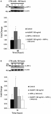
Effects of rhLIGHT, rhIFN-γ, and rhLIGHT + rhIFN-γ on the abundance of cIAP-2 protein in CTB cells at 48 hours (A) and 96 hours (B). CTB cells were incubated with (left to right) control PBS (C), rhLIGHT (100 ng/ml) (L), rhIFN-γ (100 U/ml) (I), or rhLIGHT (100 ng/ml) + rhIFN-γ (100 U/ml) (L/I), then immunoblotted to reveal cIAP-2 protein. The blots were then incubated to reveal actin. Arrows indicate cIAP-2 and actin protein, respectively. Data gathered by scanning densitometer representing the mean fold change over control (normalized to actin) ± SEM of three preparations of CTB cells, purified from three different placentas, are shown below representative blots.
Figure 5
Figure 5
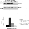
Immunoblot of protein from HT-29 cells treated for 96 hours with control PBS (C), rhLIGHT (100 ng/ml) (L), rhIFN-γ (10 U/ml) (I), or rhLIGHT (100 ng/ml) + rhIFN-γ (10 U/ml) (L/I), for cIAP-2 (and actin). Arrows indicate cIAP-2 and actin protein, respectively. Data gathered by scanning densitometer representing the fold change over control (normalized to actin), are shown below the blot. Representative of two independent experiments.
Figure 6
Figure 6
Model system for testing cIAP-2 function. A: Imaging of fluorescein-tagged morpholinos inside HT-29 cells 72 hours after ethoxylated polyethylenimine delivery. B: Immunoblot for cIAP-2 in HT-29 cells treated for 96 hours with control morpholino [control (C)] or cIAP-2 anti-sense morpholino [test (T)]. Arrows indicate cIAP-2 or actin protein, respectively. Data gathered by scanning densitometer representing the fold change over control (normalized to actin), are shown below the blot. Representative of two independent experiments. Original magnification, ×200 (A).
Figure 7
Figure 7
MTT analysis of HT-29 cells treated first with cIAP-2 or control morpholinos and then with PBS (control) or rhLIGHT (1 to 1000 ng/ml) for 96 hours. Data are shown as a percentage of the control values (no cytokine added). Error bars represent the means ± SEM of three preparations of CTB cells purified from three different placentas. Each assay was performed in triplicate. *, P < 0.05.
Figure 8
Figure 8
A schematic illustration summarizing regulation of LIGHT/IFN-γ-mediated apoptosis in human villous CTB cells.

References

    1. Phillips TA, Ni J, Hunt JS. Death-inducing tumour necrosis factor (TNF) superfamily ligands and receptors are transcribed in human placentae, cytotrophoblasts, placental macrophages and placental cell lines. Placenta. 2001;22:663–672. - PubMed
    1. Tamada K, Shimozaki K, Chapoval AI, Zhai Y, Su J, Chen S, Hsieh S, Nagata S, Ni J, Chen L. LIGHT, a TNF-like molecule, costimulates T cell proliferation and is required for dendritic cell-mediated allogenic T cell response. J Immunol. 2000;164:4105–4110. - PubMed
    1. Morel Y, Schiano de Colella J, Harrop J, Deen KC, Holmes SD, Wattam TA, Khandejar SS, Truneh A, Sweet RW, Gastaut J, Olive D, Costello RT. Reciprocal expression of the TNF family receptor herpes virus entry mediator and its ligand LIGHT on activated T cells: LIGHT downregulates its own receptors. J Immunol. 2000;165:4397–4404. - PubMed
    1. Granger SW, Butrovich KD, Houshmand P, Edwards WR, Ware CF. Genomic characterization of LIGHT reveals linkage to an immune response locus on chromosome 19p13.3 and distinct isoforms generated by alternate splicing or proteolysis. J Immunol. 2001;167:5122–5128. - PubMed
    1. Harrop JA, McDonnell PC, Brigham-Burke M, Lyn SD, Minton J, Tan KB, Dede K, Spampanato J, Silverman C, Hensley P, DiPrinzio R, Emery JG, Deen K, Eichman C, Chabot-Fletcher M, Truneh A, Young P. Herpesvirus entry mediator ligand (HVEM-L), a novel ligand for HVEM/TR2, stimulates proliferation of T-cells and inhibits HT29 cell growth. J Biol Chem. 1998;273:27548–27556. - PubMed

Publication types

MeSH terms