. 2004 Jun 23;24(25):5816-26.
doi: 10.1523/JNEUROSCI.1184-04.2004.

The GDP-GTP exchange factor collybistin: an essential determinant of neuronal gephyrin clustering

Affiliations

The GDP-GTP exchange factor collybistin: an essential determinant of neuronal gephyrin clustering

Kirsten Harvey et al. J Neurosci. .

Abstract

Glycine receptors (GlyRs) and specific subtypes of GABA(A) receptors are clustered at synapses by the multidomain protein gephyrin, which in turn is translocated to the cell membrane by the GDP-GTP exchange factor collybistin. We report the characterization of several new variants of collybistin, which are created by alternative splicing of exons encoding an N-terminal src homology 3 (SH3) domain and three alternate C termini (CB1, CB2, and CB3). The presence of the SH3 domain negatively regulates the ability of collybistin to translocate gephyrin to submembrane microaggregates in transfected mammalian cells. Because the majority of native collybistin isoforms appear to harbor the SH3 domain, this suggests that collybistin activity may be regulated by protein-protein interactions at the SH3 domain. We localized the binding sites for collybistin and the GlyR beta subunit to the C-terminal MoeA homology domain of gephyrin and show that multimerization of this domain is required for collybistin-gephyrin and GlyR-gephyrin interactions. We also demonstrate that gephyrin clustering in recombinant systems and cultured neurons requires both collybistin-gephyrin interactions and an intact collybistin pleckstrin homology domain. The vital importance of collybistin for inhibitory synaptogenesis is underlined by the discovery of a mutation (G55A) in exon 2 of the human collybistin gene (ARHGEF9) in a patient with clinical symptoms of both hyperekplexia and epilepsy. The clinical manifestation of this collybistin missense mutation may result, at least in part, from mislocalization of gephyrin and a major GABA(A) receptor subtype.


Figures

Figure 1.
Figure 1.
Alternative splicing of rat and human collybistin mRNAs. Three different collybistin C termini can be specified in the rat by the use of different cassette exons. For example, the CB2 (VTQRKWHY*) C terminus is generated by insertion of a 62 bp exon (A, white lettering on black background). When omitted, an alternative C terminus is specified (CB3) that is virtually identical to the C terminus of human collybistin (hPEM-2). RT-PCR analysis shows that the exon encoding the N-terminal SH3 domain (B) is used in most rodent and human collybistin mRNAs (○, SH3+; □, SH3-). For C-terminal exons (C), both CB2 (○) and CB3 (□) are common in rodents, but only CB3 transcripts are evident in humans. D, Schematic diagram of the primary structures of known collybistin isoforms. Sequences encoding the SH3, RhoGEF, and PH domains in the collybistin isoforms are indicated by grayboxes, and the different C termini are indicated by white (CB1), black (CB2), or gray (CB3) boxes. E, Expression of myc-tagged CB2SH3- in HEK293 cells. F, G, Cotransfection of a plasmid construct encoding EGFP-gephyrin results in the formation of cytoplasmic aggregates (F) to which myc-CB2SH3+ targets (G). However, coexpression of EGFP-gephyrin with the CB2 variant lacking the SH3 domain (myc-CB2SH3-) results in a redistribution of EGFP-gephyrin and collybistin to submembrane microaggregates (H, I). Scale bar, 10 μm. Note that E-G are single Z-plane confocal images, whereas H and I are Z-stacks at the cell surface.
Figure 2.
Figure 2.
A DsRed-GlyR β fusion protein is efficiently targeted to EGFP-gephyrin aggregates in HEK293 cells. Z-stack confocal images showing triple transfection of HEK293 cells with plasmid constructs encoding a DsRed-GlyR β subunit intracellular loop fusion protein (A; DsRed-GlyR β), EGFP-gephyrin (B), and myc-CB2SH3- (C). Note that DsRed-GlyR β is readily trapped by EGFP-gephyrin submembrane clusters, whereas myc-CB2SH3- has a more diffuse distribution pattern throughout the cytoplasm (D). Scale bar, 10 μm.
Figure 3.
Figure 3.
Functional collybistin is required for accumulation of gephyrin in dendritic clusters. A-E, Expression of myc-tagged CB2SH3- (A, C) and CB2SH3+ (B, D) isoforms in transfected mouse cortical neurons reveals a diffuse distribution throughout the cell soma and dendrites (see enlargements in C and D) without affecting gephyrin cluster number (E; values are mean ± SE). Constructs lacking the RhoGEF or PH domains (inset) were coexpressed with EGFP-gephyrin in HEK293 cells. F, G, Single Z-plane confocal images show that myc-CB2SH3-Δ RhoGEF mutant no longer colocalizes with EGFP-gephyrin and prevents submembrane targeting. H, I, In contrast, the myc-CB2SH3-Δ PH mutant colocalizes with EGFP-gephyrin but also results in the production of cytoplasmic collybistin-gephyrin microclusters. Note that gephyrin and collybistin aggregates in F-I are intracellular, because the cell nucleus is clearly visible in each single optical section. J, K, In transfected mouse cortical neurons, myc-CB2SH3-Δ PH acts as a dominant-negative factor, competing with endogenous collybistin for binding sites on gephyrin. Quantitative analysis of mAb7a staining indicates a statistically significant loss of dendritic gephyrin clusters (E;*p < 0.001; Student's t test; n = 10-11 cells per construct) and accumulation of endogenous gephyrin in aggregates in the proximal dendrites (white arrow). Scale bars: A, B, 22 μm; C, D, 11 μm; F-I, 5 μm; J, 23 μm; K, 11.5 μm.
Figure 4.
Figure 4.
Mapping of the collybistin binding site on gephyrin. A, Deletion constructs of the rat gephyrin P1 isoform in pACT2 (Clontech) were tested using the GlyR β subunit intracellular loop (β) and full-length collybistin CB2SH3- isoform (CB2) as baits. Both proteins interact with full-length gephyrin in yeast, as judged by a semiquantitative LacZ assay (+++, strong interaction; +, weak interaction; —, no interaction). Deletion analysis indicated that the gephyrin MogA domain (MogA) and most of the linker region do not appear to contribute to either GlyR β subunit or collybistin binding. Deletions into the gephyrin MoeA domain (MoeA) destroy interactions with both collybistin and the GlyR β subunit because of the loss of MoeA-MoeA domain interactions. B, Geph-305 interacts with GlyR β, CB2SH3-, and MoeA domain baits, whereas Geph-323 (which corresponds to a MoeA domain prey) interacts with GlyR β and MoeA but not CB2SH3-. C, D, Mutants (Geph-A1 to Geph-A7), which span the Geph-305 to Geph 323 region, were then constructed and tested in yeast assays. Mutations Geph-A4 and Geph-A5 (boxed) abolished interactions with CB2SH3- but not the GlyR β subunit (C, D). The corresponding mutants in EGFP-gephyrin eliminated (EGFP-GephA4) or weakened (EGFP-GephA5) colocalization with myc-CB2SH3- (E) and blocked submembrane microaggregate formation. Scale bar, 10 μm.
Figure 5.
Figure 5.
Disruption of the collybistin binding site on gephyrin prevents accumulation of gephyrin at postsynaptic sites. A-H, EGFP-tagged gephyrin (A, E) and the mutants EGFP-GephA4 (B, C, F, G) and EGFP-GephA5 (D, H) were transfected into cortical neurons and the cells immunostained for gephyrin (mAb7a; A-D) or GAD (mAbGAD-6; E-H). Two examples of cells are shown for EGFP-GephA4 representing comparatively low (B, F) and high (C, G) levels of expression and corresponding aggregate phenotypes. Note the perfect colocalization of transfected EGFP-gephyrin with endogenous gephyrin in yellow puncta (A). The few remaining red puncta indicate endogenous gephyrin expressed on dendrites of nontransfected neighboring cells that are therefore not colocalized with EGFP-gephyrin. EGFP-gephyrin is localized at postsynaptic sites juxtaposed to presynaptic GAD (E, arrows). Mutants EGFP-GephA4 and EGFP-GephA5 form large aggregates in the soma and dendrites, which are no longer juxtaposed to GABAergic terminals (F-H). Endogenous gephyrin has been trapped in these extrasynaptic gephyrin mutant aggregates, which therefore appear yellow (B-D). Quantitative analysis of mAb7a staining indicates a statistically significant loss of synaptic gephyrin clusters (I;*p < 0.001; Student's t test; n = 12-13 cells per construct) for the EGFP-GephA4 and EGFP-GephA5 mutants compared with EGFP-gephyrin. Scale bar, 20 μm.
Figure 6.
Figure 6.
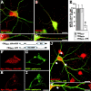
Mutation of G55A in a patient with hyperekplexia and epilepsy. A, Genomic structure of the human collybistin gene (ARHGEF9) encoding hPEM-2. B, The location of the mutation G55A (red lettering) in the N-terminal SH3 domain (underlined) of hPEM-2 is shown. G55A lies proximal to one of the series of residues that have been proposed to bind proline-rich motifs (blue lettering). C, DNA sequencing of exon 2 samples revealed a G to C substitution at position 164 of the collybistin coding sequence, causing a G55A missense mutation in the N-terminal SH3 domain of hPEM2. G164, Control sample; G164+G164C, ad-mixture of the patient sample with a female DNA control; G164C, G164C mutation. D, Restriction fragment length polymorphism analysis of the G55A mutation: the G164C mutation creates a novel NlaIII site at position 207 of the 313 bp exon 2 PCR fragment. This, in conjunction with another NlaIII site at position 106, generates convergent fragments of 106, 101, and 106 bp in the patient DNA sample (denoted P). Normal controls (C1-C4) have fragments of 207 and 106 bp. E, A molecular model showing the location of G55 in the SH3 domain structure calculated with Swiss-Model (Guex and Peitsch, 1997). F, G, Confocal images showing HEK293 cells transfected with plasmid constructs for myc-CB3SH3+ and EGFP-gephyrin (F) or myc-CB3SH3+G55A and EGFP-gephyrin (G). Note that myc-CB3SH3+colocalizes with cytoplasmic EGFP-gephyrin aggregates, whereas myc-CB3SH3+G55A results in submembrane microclusters of EGFP-gephyrin. H-P, Confocal images showing myc-CB3SH3+ (H-J) and myc-CB3SH3+G55A (K-P) expressed in cultured cortical neurons. Although wild-type myc-CB3SH3+ (H) is expressed throughout the cell soma and dendrites and does not disrupt gephyrin localization (H-J), myc-CB3SH3+G55A forms a tight association with endogenous gephyrin in clusters confined to proximal dendrites (K-M). In a subset of transfected cells (N-P), myc-CB3SH3+G55A forms large somatic and dendritic aggregates, which results in an almost complete loss of gephyrin clusters. Remaining red puncta in I, L, and O represent gephyrin clusters from nontransfected cells in the same culture. Quantitative analysis of mAb7a immunofluorescence (Q) indicates a dramatic loss of gephyrin clusters (*p < 0.001; Student's t test; n = 11 cells per construct) for myc-CB3SH3+G55A mutant compared with myc-CB3SH3+. Scale bars: F, G, 10 μm; H-P, 35 μm.
Figure 7.
Figure 7.
Collybistin mutant CB3SH3+G55A results in loss of GABAA receptor clusters. Triple staining of neurons transfected with either myc-CB3SH3+ (A-H) or myc-CB3SH3+G55A (I-P) with antibodies against the GABAA receptor γ2 subunits (red), myc tag (green), and GAD (blue). Note that in myc-CB3SH3+-transfected neurons (A-D), GABAA receptor clusters are juxtaposed to GAD-positive terminals (a selected dendritic region is enlarged in images E-H). Overlap of GABAA receptor and GAD immunoreactivity appears pink in the merged image (H, arrows). In contrast, in neurons expressing myc-CB3SH3+G55A, GABAA receptor immunoreactivity is primarily confined to the cell soma (I, M), and large collybistin aggregates (J, N) are observed throughout the dendrites. Note that these collybistin aggregates are not juxtaposed to GABAergic terminals (K, L, O, P). Quantitative analysis of γ2 subunit immunofluorescence (Q) indicates a statistically significant loss of GABAA receptor clusters (*p < 0.001; Student's t test; n = 12 cells per construct) for the myc-CB3SH3+G55A mutant compared with myc-CB3SH3+. Scale bars: A-D, I-L, 25 μm; E-H, M-P, 12.5 μm.

References

    1. Andrew M, Owen MJ (1997) Hyperekplexia: abnormal startle response due to glycine receptor mutations. Br J Psychiatry 170: 106-108. - PubMed
    1. Bagrodia S, Taylor SJ, Jordon KA, Van Aelst L, Cerione RA (1998) A novel regulator of p21-activated kinases. J Biol Chem 273: 23633-23636. - PubMed
    1. Douangamath A, Filipp FV, Klein AT, Barnett P, Zou P, Voorn-Brouwer T, Vega MC, Mayans OM, Sattler M, Distel B, Wilmanns M (2002) Topography for independent binding of alpha-helical and PPII-helical ligands to a peroxisomal SH3 domain. Mol Cell 10: 1007-1017. - PMC - PubMed
    1. Dunne EL, Hosie AM, Wooltorton JR, Duguid IC, Harvey K, Moss SJ, Harvey RJ, Smart TG (2002) An N-terminal histidine regulates Zn2+ inhibition on the murine GABAA receptor β3 subunit. Br J Pharmacol 137: 29-38. - PMC - PubMed
    1. Erickson JW, Cerione RA (2001) Multiple roles for Cdc42 in cell regulation. Curr Opin Cell Biol 13: 153-157. - PubMed

Publication types

MeSH terms