. 2004 Aug;15(8):3950-63.
doi: 10.1091/mbc.e04-01-0019. Epub 2004 Jun 23.

Promotion of neurite and filopodium formation by CD47: roles of integrins, Rac, and Cdc42

Affiliations

Promotion of neurite and filopodium formation by CD47: roles of integrins, Rac, and Cdc42

Motoaki Miyashita et al. Mol Biol Cell. 2004 Aug.

Abstract

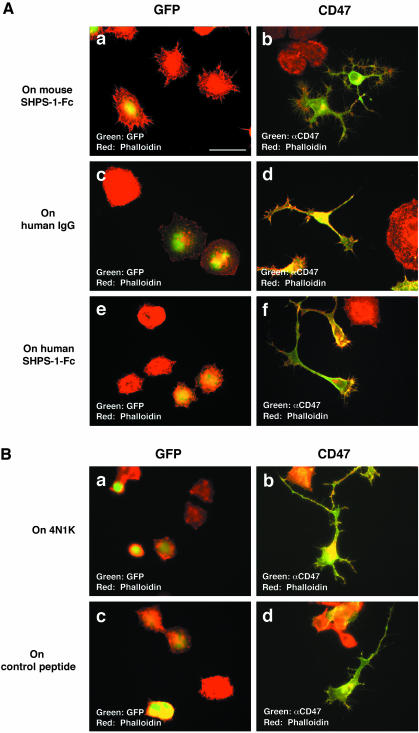
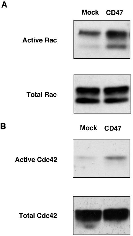
Axon extension during development is guided by many factors, but the signaling mechanisms responsible for its regulation remain largely unknown. We have now investigated the role of the transmembrane protein CD47 in this process in N1E-115 neuroblastoma cells. Forced expression of CD47 induced the formation of neurites and filopodia. Furthermore, an Fc fusion protein containing the extracellular region of the CD47 ligand SHPS-1 induced filopodium formation, and this effect was enhanced by CD47 overexpression. SHPS-1-Fc also promoted neurite and filopodium formation triggered by serum deprivation. Inhibition of Rac or Cdc42 preferentially blocked CD47-induced formation of neurites and filopodia, respectively. Overexpression of CD47 resulted in the activation of both Rac and Cdc42. The extracellular region of CD47 was sufficient for the induction of neurite formation by forced expression, but the entire structure of CD47 was required for enhancement of filopodium formation by SHPS-1-Fc. Neurite formation induced by CD47 was also inhibited by a mAb to the integrin beta3 subunit. These results indicate that the interaction of SHPS-1 with CD47 promotes neurite and filopodium formation through the activation of Rac and Cdc42, and that integrins containing the beta3 subunit participate in the effect of CD47 on neurite formation.


Figures

Figure 1.
Figure 1.
Expression of CD47 and effects of serum deprivation on neurite formation in N1E-115 cells. (A) Lysates of N1E-115 cells, of CHO-Ras cells, or of CHO-Ras cells stably expressing mouse CD47 form 2 (CHO-Ras-CD47) were subjected to immunoblot analysis with a mAb to CD47 (αCD47). (B) N1E-115 cells transfected with a vector for GFP (Mock) or with a vector containing mouse CD47 form 2 or form 4 cDNA were lysed 24 h after transfection and subjected to immunoblot analysis with a mAb to CD47. (C) N1E-115 cells were cultured in the absence (Serum (-); a and c) or presence (Serum (+); b and d) of 10% FBS for 24 h, fixed, and stained with a mAb to CD47 (c and d); phase-contrast images of the same cells are also shown (Phase; a and b). Scale bar, 50 μm. All results shown are representative of three separate experiments.
Figure 2.
Figure 2.
Neurite and filopodium formation induced in N1E-115 cells by overexpression of CD47. (A) N1E-115 cells were transfected with expression vectors for CD47 form 2 (a and d), CD47 form 4 (b and e), or GFP (c and f). After incubation for 24 h in the presence of 10% FBS, the cells were stained with rhodamine-conjugated phalloidin (Phalloidin; a-c) or a mAb to CD47 (d and e); GFP fluorescence (GFP) is shown in f. Arrows indicate cells expressing recombinant CD47 or GFP. Scale bar, 50 μm. (B) N1E-115 cells were cotransfected with vectors for GFP and CD47 form 2 or 4 (or the corresponding empty vector [Vector]). After incubation for 24 h in the presence of 10% FBS, the cells were stained with a mAb to CD47. The percentage of cells expressing both CD47 and GFP that exhibited neurites at least as long as the cell body was determined. The effect of serum deprivation on neurite formation was also quantified for N1E-115 cells transfected with the GFP vector alone (Serum (-)). Data are means ± SE of values from three separate experiments. (C) N1E-115 cells were transfected with an expression vector for GFP (a and c) or for CD47 form 4 (b). After culture for 24 h in the presence (a and b) or absence (c) of 10% FBS, the cells were stained with rhodamine-conjugated phalloidin (red). Expression of GFP or CD47 was detected by GFP fluorescence and immunostaining with a mAb to CD47, respectively (green). Scale bar, 10 μm. Results in A and C are representative of three separate experiments.
Figure 3.
Figure 3.
Effects of an SHPS-1–Fc fusion protein and forced expression of CD47 on filopodium formation. (A) N1E-115 cells plated on dishes coated with mouse SHPS-1–Fc (a and b), control human IgG (c and d), or human SHPS-1–Fc (e and f) were transfected with a vector for either GFP (a, c, and e) or CD47 form 4 (b, d, and f). After culture for 24 h in the presence of 10% FBS, the cells were stained with rhodamine-conjugated phalloidin (red); those transfected with the CD47 vector were also immunostained with a mAb to CD47 (green), whereas those transfected with the GFP vector were detected by GFP fluorescence (green). (B) N1E-115 cells plated on dishes coated with the 4N1K peptide (a and b) or a control peptide (c and d) were transfected with a vector for either GFP (a and c) or CD47 form 4 (b and d). After culture for 24 h in the presence of 10% FBS, the cells were stained as in A. Scale bar, 50 μm. All results are representative of three separate experiments.
Figure 4.
Figure 4.
Promotion by mouse SHPS-1–Fc of neurite and filopodium formation triggered in N1E-115 cells by serum deprivation. (A) N1E-115 cells were plated on dishes coated with control human IgG (a-c) or with mouse SHPS-1–Fc (d-f) and were cultured for 5 h in the presence of 10% FBS. After incubation in the absence of serum for an additional 1 h (a and d),3h(bande),or15h(candf),the cells were stained with rhodamine-phalloidin. Scale bar, 50 μm. (B) Cells were plated on dishes coated with control human IgG (a) or with mouse SHPS-1–Fc (b) and subjected to serum deprivation for 1 h as in A before staining with rhodamine-phalloidin. Scale bar, 10 μm. (C) Cells treated and analyzed as in A were evaluated for neurite extension. The percentages of cells with neurites whose length corresponded to 1× to 2× (open column), 2× to 3× (gray column), or >3× (closed column) that of the cell body were determined. Data are means ± SE of three separate experiments. All results are representative of three separate experiments.
Figure 5.
Figure 5.
Effects of a dominant negative mutant of Rac and of NWASP-CRIB on CD47-dependent neurite and filopodium formation. (A) N1E-115 cells were cotransfected with a vector for CD47 form 4 and a vector for either GFP (GFP + CD47; a and d), GFP-N17Rac1 (GFP-RacDN; GFP-RacDN + CD47; b and e), or GFP-NWASP–CRIB (GFP-NWASP-CRIB + CD47; c and f). After culture for 24 h in the presence of 10% FBS, the cells were immunostained with a mAb to CD47 (d-f) or monitored for GFP fluorescence (a-c). Scale bar, 50 μm. (B) N1E-115 cells were cotransfected with a vector for CD47 form 4 (closed column; or the corresponding empty vector; open column) and a vector for either GFP (GFP + vector, GFP + CD47), GFP-N17Rac1 (RacDN + vector, RacDN + CD47), or GFP-NWASP–CRIB (NWASP-CRIB + vector, NWASP-CRIB + CD47). After culture for 24 h in the presence of 10% FBS, the cells were analyzed as in A and neurite formation was quantitated as described in Figure 2B. Data are means ± SE of values from three separate experiments. (C) N1E-115 cells were plated on dishes coated with mouse SHPS-1–Fc and were transfected, cultured, and analyzed as in A. Scale bar, 50 μm. Results in A and C are representative of three separate experiments.
Figure 6.
Figure 6.
Activation of Rac and Cdc42 by forced expression of CD47. COS-7 cells were transfected with a vector for CD47 form 4 (CD47) or the corresponding empty vector (Mock). The cells were lysed 24 h after transfection, and the GTP-bound (active) forms of Rac (A) or Cdc42 (B) were precipitated with a GST fusion protein containing the Rac/Cdc42 binding domain of p21PAKα. The resulting precipitates were subjected to immunoblot analysis with mAbs to Rac or to Cdc42 (top panels). Whole cell lysates were also directly subjected to immunoblot analysis with the same mAbs to determine the total amounts of Rac or Cdc42 (bottom panels). Results are representative of three separate experiments.
Figure 7.
Figure 7.
Identification of the regions of CD47 responsible for the promotion of neurite and filopodium formation. (A) Schematic representation of the CD47 mutants studied. CD47WT, wild-type CD47 form 4. (B) N1E-115 cells were transfected with a vector for CD47WT (a), CD47EX-TM (b), or CD8-CD47MMS (c), incubated for 24 h in the presence of 10% FBS, and stained with rhodamine-phalloidin (red; a-c) as well as with mAbs to either CD47 (green; a and b) or CD8 (green; c). Scale bar, 50 μm. (C) N1E-115 cells were cotransfected with a vector for GFP and the vectors for indicated mutants (or the corresponding empty vector [Vector]), incubated for 24 h in the presence of 10% FBS, and then stained with a mAb to CD47. Quantitative analysis of neurite formation was performed as in Figure 2B. Data are means ± SE of values from three separate experiments. (D) N1E-115 cells plated on dishes coated with mouse SHPS-1–Fc were transfected, incubated, and stained as in A. Scale bar, 50 μm. Results in B and D are representative of three separate experiments.
Figure 8.
Figure 8.
Effects of echistatin, inhibitory mAbs to integrin subunits, PTX, or wortmannin on CD47-induced neurite and filopodium formation. (A) N1E-115 cells were cotransfected with a vector for CD47 (closed column; or the corresponding empty vector; open column) and a vector for GFP and were then cultured for 24 h with 10% FBS in the absence (Control) or presence of echistatin (0.05 μg/ml). Alternatively, the transfected cells were cultured for 24 h, detached from the dish, incubated for 5 min with normal mouse IgG (5 μg/ml; Mouse IgG) or with mAbs to the integrin β3 (5 μg/ml; β3-antibody) or β1 (5 μg/ml; β1-antibody) subunits, and then replated and cultured for 24 h. All cells were then visualized either by CD47 immunofluorescence or by GFP fluorescence, and neurite formation was quantified as in Figure 2B. Data are means ± SE of values from three separate experiments. (B) N1E-115 cells plated on dishes coated with mouse SHPS-1–Fc were transfected with a vector for CD47 and treated with echistatin or the mAbs to integrin β1 or β3 subunits as in A. They were then stained with rhodamine-phalloidin (red) and a mAb to CD47 (green). Scale bar, 50 μm. (C) N1E-115 cells were plated on dishes coated with control human IgG or with mouse SHPS-1–Fc and were cultured for 4 h in the presence of 10% FBS. Cells were subjected to serum deprivation in the absence or presence of echistatin (0.05 μg/ml). After incubation for 1 h, the percentages of cells with neurites whose length corresponded to 1× to 2× (open column), 2× to 3× (gray column), or >3× (closed column) that of the cell body were determined. Data are means ± SE of values obtained from 15 randomly chosen fields. (D) N1E-115 cells were cotransfected with a vector for CD47 (closed column; or the corresponding empty vector; open column) and a vector for GFP, cultured for 24 h with 10% FBS in the absence (Control) or presence of PTX (100 ng/ml) or wortmannin (100 nM), and then stained with a mAb to CD47. Quantitative analysis of neurite formation was performed as in Figure 2B. Data are means ± SE of values from three separate experiments. (E) N1E-115 cells plated on dishes coated with mouse SHPS-1–Fc were transfected with a vector for CD47, cultured for 24 h with 10% FBS in the absence (Control; a) or presence of PTX (100 ng/ml; b), or wortmannin (100 nM; c), and stained with rhodamine-phalloidin (red) and a mAb to CD47 (green). Scale bar, 50 μm. Results in B, C, and E are representative of three separate experiments.
Figure 9.
Figure 9.
Effects of an SHPS-1–Fc fusion protein and forced expression of CD47 on filopodium formation in cultured hippocampal neurons. Mouse hippocampal neurons were cultured on dishes coated with control human IgG (A and B) or mouse SHPS-1-Fc (C and D) for 3 days. Neurons were then cotransfected with the vectors for GFP-actin and CD47 (A-D). Four days after transfection, neurons were immunostained with a mAb to CD47 (red). The cell morphology was examined by fluorescence of GFP-actin (green). Magnification: (A and C) ×630; (B and D) ×1000. Scale bar for A, 20 μm; for B, 10 μm. All results are representative of three separate experiments.

References

    1. Arakawa, Y., Bito, H., Furuyashiki, T., Tsuji, T., Takemoto-Kimura, S., Kimura, K., Nozaki, K., Hashimoto, N., and Narumiya, S. (2003). Control of axon elongation via an SDF-1α/Rho/mDia pathway in cultured cerebellar granule neurons. J. Cell Biol. 161, 381-391. - PMC - PubMed
    1. Brown, E.J., and Frazier, W.A. (2001). Integrin-associated protein (CD47) and its ligands. Trends Cell Biol. 11, 130-135. - PubMed
    1. Brown, E.J., Hooper, L., Ho, T., and Gresham, H. (1990). Integrin-associated protein: a 50-kD plasma membrane antigen physically and functionally associated with integrins. J. Cell Biol. 111, 2785-2794. - PMC - PubMed
    1. Chang, H.P., Lindberg, F.P., Wang, H.L., Huang, A.M., and Lee, E.H. (1999). Impaired memory retention and decreased long-term potentiation in integrin-associated protein-deficient mice. Learn. Mem. 6, 448-457. - PMC - PubMed
    1. Chung, J., Gao, A.G., and Frazier, W.A. (1997). Thrombospondin acts via integrin-associated protein to activate the platelet integrin αIIbβ3. J. Biol. Chem. 272, 14740-14746. - PubMed

Publication types

MeSH terms