. 2004 Sep;15(9):3994-4002.
doi: 10.1091/mbc.e04-01-0053. Epub 2004 Jun 23.

A type V myosin (Myo2p) and a Rab-like G-protein (Ypt11p) are required for retention of newly inherited mitochondria in yeast cells during cell division

Affiliations

A type V myosin (Myo2p) and a Rab-like G-protein (Ypt11p) are required for retention of newly inherited mitochondria in yeast cells during cell division

Istvan R Boldogh et al. Mol Biol Cell. 2004 Sep.

Abstract

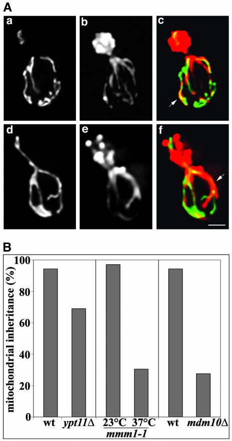
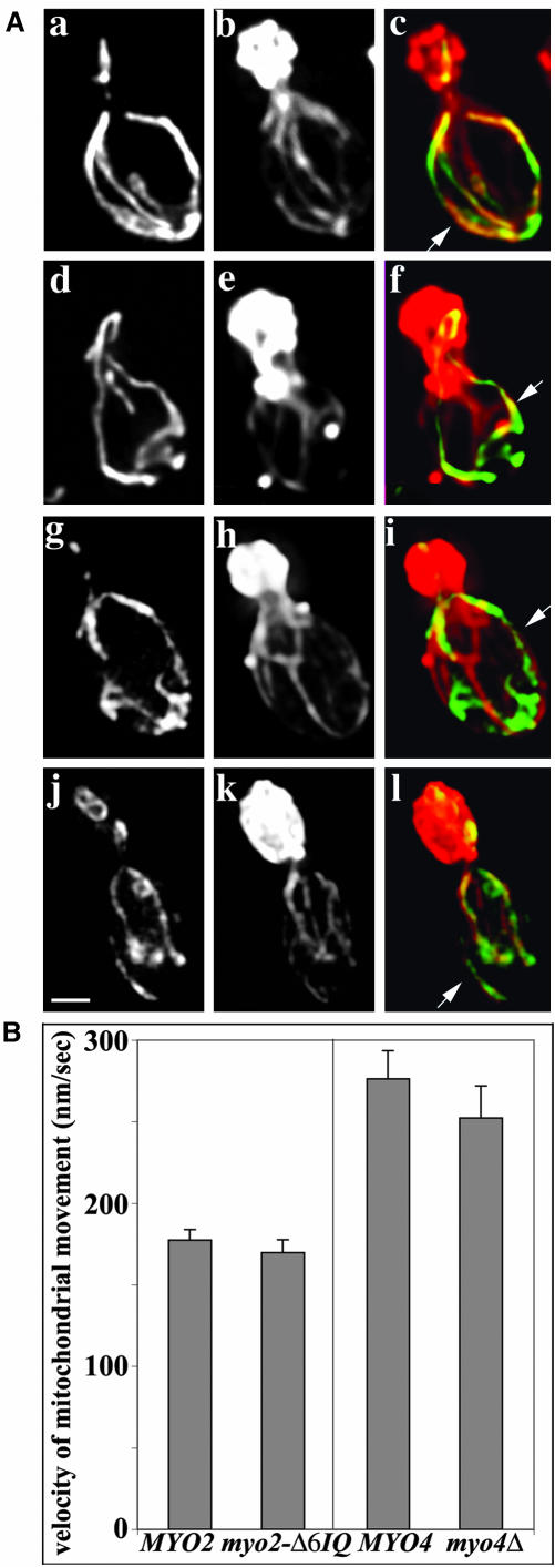
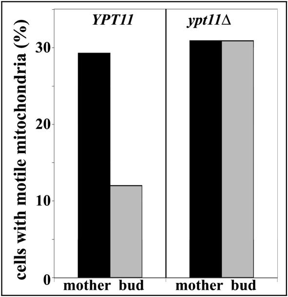
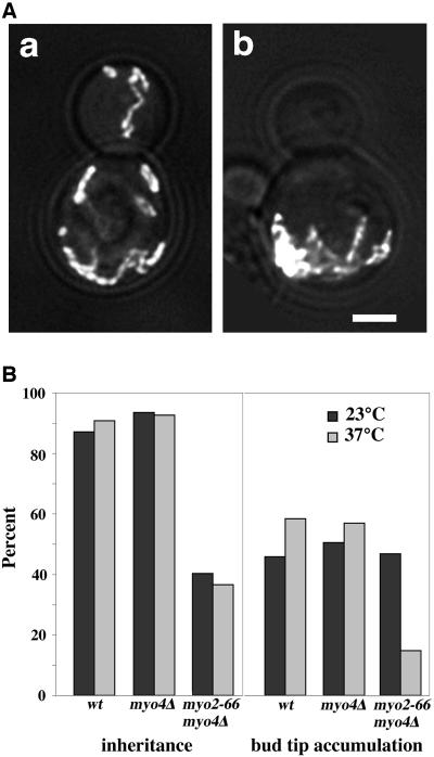
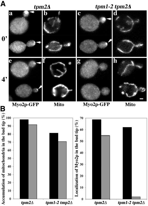
Two actin-dependent force generators contribute to mitochondrial inheritance: Arp2/3 complex and the myosin V Myo2p (together with its Rab-like binding partner Ypt11p). We found that deletion of YPT11, reduction of the length of the Myo2p lever arm (myo2-Delta6IQ), or deletion of MYO4 (the other yeast myosin V), had no effect on mitochondrial morphology, colocalization of mitochondria with actin cables, or the velocity of bud-directed mitochondrial movement. In contrast, retention of mitochondria in the bud was compromised in YPT11 and MYO2 mutants. Retention of mitochondria in the bud tip of wild-type cells results in a 60% decrease in mitochondrial movement in buds compared with mother cells. In ypt11Delta mutants, however, the level of mitochondrial motility in buds was similar to that observed in mother cells. Moreover, the myo2-66 mutant, which carries a temperature-sensitive mutation in the Myo2p motor domain, exhibited a 55% decrease in accumulation of mitochondria in the bud tip, and an increase in accumulation of mitochondria at the retention site in the mother cell after shift to restrictive temperatures. Finally, destabilization of actin cables and the resulting delocalization of Myo2p from the bud tip had no significant effect on the accumulation of mitochondria in the bud tip.


Figures

Figure 1.
Figure 1.
Deletion of YPT11 impairs mitochondrial inheritance without affecting mitochondrial morphology or interactions of mitochondria with actin cables. (A) YPT11 wild-type cells (BY4741) (a-c) and deletion mutants (RG1140) (d-f) expressing mitochondria-targeted GFP (CS1-GFP) were grown to mid-log phase. Cells were fixed and stained with rhodamine-phalloidin. z-Sections of cells were collected, deconvolved, and projected to a single image. GFP-labeled mitochondria (a and d), actin organization (b and e), as well as an overlay of mitochondria in green and actin in red (c and f) are shown. Arrows point to examples of colocalization of mitochondria with actin cables. Bar, 1 μm. (B) YPT11 wild-type cells (BY4741) and deletion mutants (RG1140), and MDM10 wild-type cells (MYY291) and deletion mutants (MYY504) were grown to mid-log phase. Temperature-sensitive mmm1-1 mutant (YPH253) cells were grown to mid-log phase at 23°C and then shifted to 37°C for 3 h and 45 min. Mitochondria were visualized in YPT11 wild type and deletion strains by using CS1-GFP. In all other strains mitochondria were visualized using the membrane potential-sensing dye DiOC6. Mitochondrial inheritance was determined by scoring the percentage of yeast bearing small buds that contained mitochondria within the bud.
Figure 2.
Figure 2.
Mutation of either of the type V myosins of yeast has no effect on mitochondrial morphology, association of mitochondria with actin cables, or the rate of movement of mitochondria from mother cells to buds. (A) Wild-type MYO2 (CRY1) (a-c), myo2-Δ6IQ (RSY21) cells (d-f), wild-type MYO4 (22AB) cells (g-i), and myo4Δ (MYO4ΔU5) mutants (j-l) expressing CS1-GFP were grown to midlog phase at 30°C. Cells were fixed and stained with rhodaminephalloidin. z-Sections of cells were collected, deconvolved, and projected to a single image. GFP-labeled mitochondria (a, d, g, and j), actin organization (b, e, h, and k), and the overlay of mitochondria in green and actin in red (c, f, i, and l) are shown. Arrows point to examples of colocalization of mitochondria with actin cables. Bar, 1 μm. (B) Velocities of mitochondrial movement in MYO2 wild-type cells, myo2-Δ6IQ mutant cells, MYO4 wild-type and myo4Δ cells expressing CS1-GFP were measured by time-lapse fluorescence microscopy as described under Materials and Methods.
Figure 3.
Figure 3.
Events associated with the retention of mitochondria in the bud. Wild-type yeast expressing CS1-GFP (BY4741) were grown to mid-log phase and monitored by 4D imaging (time-lapse microscopy combined with 3D reconstruction). z-Sections through the entire cell were obtained as described above. The interval between each Z-series was 5 s. The still frames shown are two-dimensional projections of deconvolved 3D reconstructions obtained at 0-25 s (top) and 155-180 s (bottom) from a single 4D time-lapse series. Arrow points to mitochondria that are immobilized and accumulated in the tip of the bud. Arrowheads in the top and bottom panels point to mitochondria that undergo linear movement toward the bud tip. Bar, 1 μm. Please refer to Supplemental Information to view a movie of this 4D time-lapse series.
Figure 4.
Figure 4.
Deletion of YPT11 results in defects in immobilization of newly inherited mitochondria in the bud. YPT11 wild-type cells (BY4741) and deletion mutants (RG1140) expressing mitochondria-targeted GFP (CS1-GFP) were grown to mid-log phase. Time-lapse fluorescence imaging was used to monitor movements of CS1-GFP-labeled mitochondria. Images were acquired at 1 s intervals over a period of 1 min. The percentage of cells displaying mitochondrial movement in mother cells and buds in wild-type and ypt11Δ cells was determined as described under Materials and Methods.
Figure 5.
Figure 5.
Mutation of the Myo2p motor domain results in defects in mitochondrial inheritance, aggregation of mitochondria in the distal tip of the mother cell, and defects in accumulation of mitochondria in the bud tip. (A) Yeast bearing mutations in both type V myosins (myo2-66 and myo4Δ; VSY21) were transformed with pCS1-GFP. The strain was grown to mid-log phase at 23°C and incubated at 23°C (a) or 37°C, the restrictive temperature for myo2-66, (b) for 45 min before fixation. z-Sections of GFP-labeled mitochondria were collected, deconvolved, and projected to a single image as described under Materials and Methods. Phase images of the same cells were overlaid on the fluorescence images. Bar, 1 μm. (B) Mid-log phase myo2-66/myo4Δ mutants (VSY21), myo4Δ mutants (MYO4ΔU5) or wild-type strains (22AB) from the same genetic background that expressed CS1-GFP, were incubated at 23 or 37°C and fixed as for A. Mitochondrial inheritance (left) was assayed as for Figure 1B. Accumulation of mitochondria in the bud tip of each cell type (right) was determined by scoring the percentage of cells that displayed a buildup of mitochondria in the tip of the bud. All cells that were analyzed for bud tip accumulation of mitochondria contained mitochondria in their bud tips.
Figure 6.
Figure 6.
Mislocalization of Myo2p has no effect on retention of mitochondria in the bud tip. (A) Yeast carrying a deletion in the tropomyosin gene TPM2 (tpm2Δ) and either wild-type tropomyosin gene (TPM1; IBY152) (a and b, e and f) or temperature-sensitive TPM1 mutation (tpm1-2; IBY153) (c and d, g and h) were grown to mid-log phase at permissive temperatures (23°C). Each of these strains expressed Myo2p tagged at its C terminus with GFP (Myo2p-GFP) and mitochondria-targeted HcRed (OLI1-HcRed). Aliquots from this culture were incubated at temperatures restrictive for the tpm1-2 allele (35°C) for 0 min (a-d) or 4 min (e-h). The cells were fixed and OLI1-HcRed-labeled mitochondria (b, d, f, and h) and Myo2p-GFP (a, c, e, and g) were visualized by fluorescence microscopy. Arrows point to instances of accumulation of Myo2p in the bud tip. Bar, 1 μm. (B) Aliquots from TPM1 tpm2Δ and tpm1-2 tpm2Δ cells were incubated at temperatures restrictive for the tpm1-2 allele (35°C) for 0 min (black bars) or 4 min (gray bars) and the cells were fixed (see above). OLI1-HcRed-labeled mitochondria (left) and Myo2p-GFP (right) were visualized by fluorescence microscopy. Accumulation of mitochondria in the bud tip was assessed as for Figure 5. Myo2p-GFP was scored as depolarized if it did not accumulate in the bud tip. Mislocalization of Myo2p had no effect on accumulation of mitochondria in the bud tip.
Figure 7.
Figure 7.
Synthetic growth defect of ypt11Δ with mdm12Δ. ypt11Δ/YPT11 mdm12Δ/MDM12 heterozygous diploid cells derived from mating the haploid strains, RG1140 (ypt11Δ) and MYY624 (mdm12Δ), were sporulated, and the tetrads were dissected. Growth characteristics of the haploid spores were tested on glucose-based solid media (YPD) and glycerol-based solid media (YPG). The panel shows a dilution series of a representative set of tetrads after 4 d of incubation at 23 and 37°C.

References

    1. Amberg, D.C., Zahner, J.E., Mulholland, J.W., Pringle, J.R., and Botstein, D. (1997). Aip3p/Bud6p, a yeast actin-interacting protein that is involved in morphogenesis and the selection of bipolar budding sites. Mol. Biol. Cell. 8, 729-753. - PMC - PubMed
    1. Beach, D.L., Thibodeaux, J., Maddox, P., Yeh, E., and Bloom, K. (2000). The role of the proteins Kar9 and Myo2 in orienting the mitotic spindle of budding yeast. Curr. Biol. 10, 1497-1506. - PubMed
    1. Boldogh, I.R., Nowakowski, D.W., Yang, H-C., Chung, H., Karmon, S., Royes, P., and Pon, L.A. (2003). A protein complex containing Mdm10p, Mdm12p and Mmm1p links mitochondrial membranes and DNA to the cytoskeleton-based segregation machinery. Mol. Biol. Cell. 14, 4618-4627. - PMC - PubMed
    1. Boldogh, I.R., Yang, H.-C., Nowakowski, W.D., Karmon, S.L., Hays, L.G., Yates, J.R., III, and Pon, L.A. (2001). Arp2/3 complex and actin dynamics are required for actin-based mitochondrial motility in yeast. Proc. Natl. Acad. Sci. USA 98, 3162-3167. - PMC - PubMed
    1. Brockerhoff, S.E., and Davis, T.N. (1992). Calmodulin concentrates at regions of cell growth in Saccharomyces cerevisiae. J. Cell Biol. 118, 619-629. - PMC - PubMed

Publication types

MeSH terms