Comparative Study
. 2004 Sep;15(9):4148-65.
doi: 10.1091/mbc.e04-01-0058. Epub 2004 Jun 23.

The basolateral targeting signal of CD147 (EMMPRIN) consists of a single leucine and is not recognized by retinal pigment epithelium

Affiliations
Comparative Study

The basolateral targeting signal of CD147 (EMMPRIN) consists of a single leucine and is not recognized by retinal pigment epithelium

Ami A Deora et al. Mol Biol Cell. 2004 Sep.

Abstract

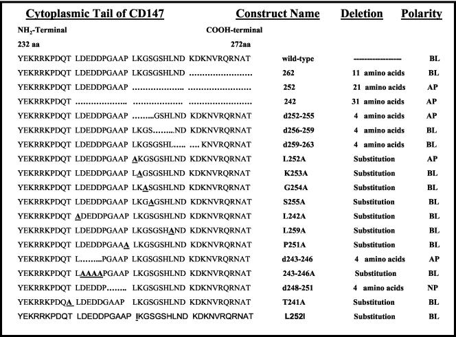
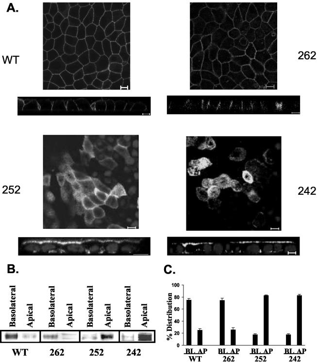
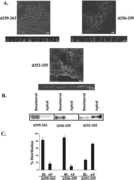
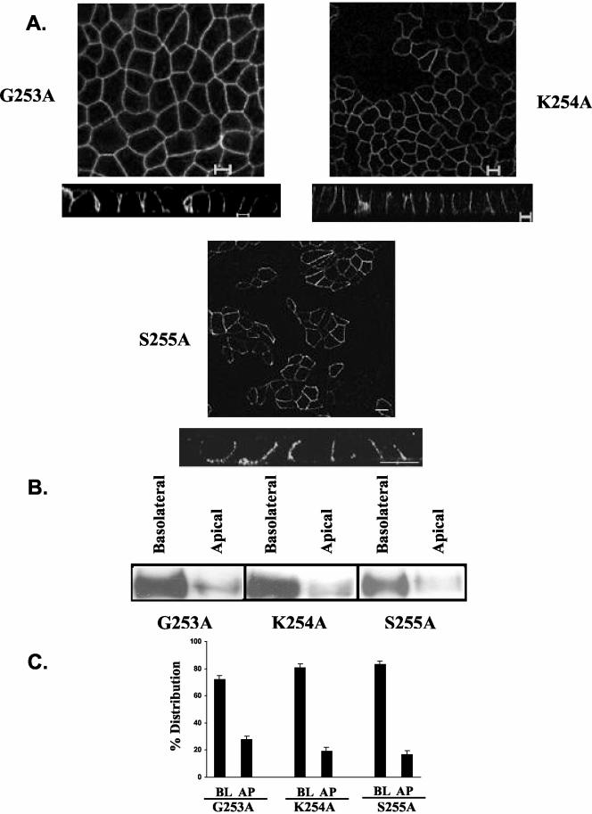
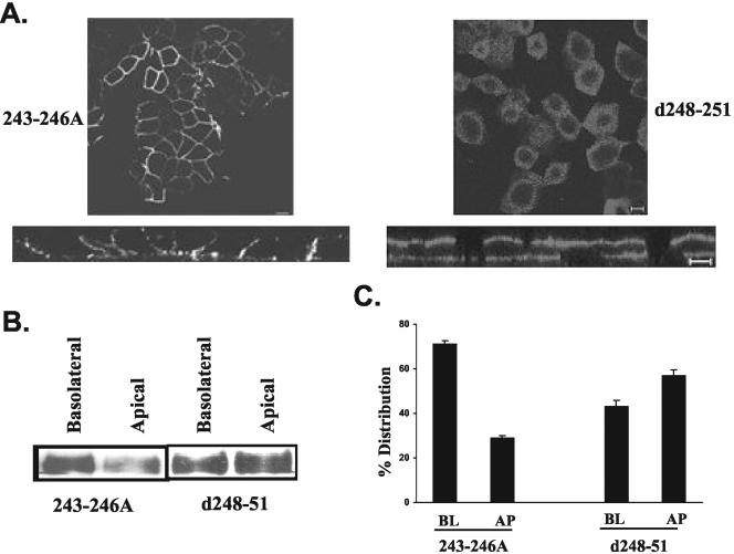
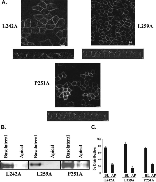
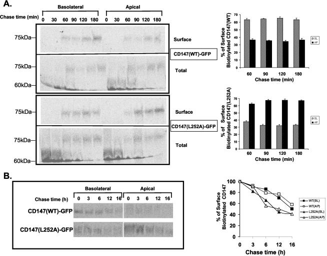
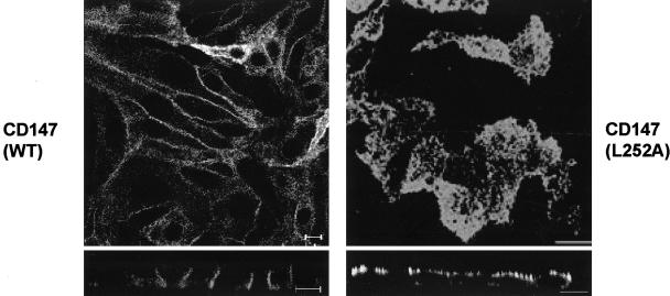
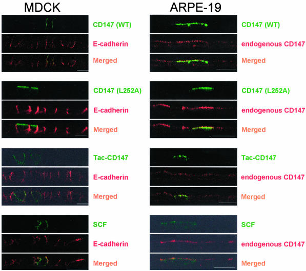
CD147, a type I integral membrane protein of the immunoglobulin superfamily, exhibits reversed polarity in retinal pigment epithelium (RPE). CD147 is apical in RPE in contrast to its basolateral localization in extraocular epithelia. This elicited our interest in understanding the basolateral sorting signals of CD147 in prototypic Madin-Darby canine kidney (MDCK) cells. The cytoplasmic domain of CD147 has basolateral sorting information but is devoid of well-characterized basolateral signals, such as tyrosine and di-leucine motifs. Hence, we carried out systematic site-directed mutagenesis to delineate basolateral targeting information in CD147. Our detailed analysis identified a single leucine (252) as the basolateral targeting motif in the cytoplasmic tail of CD147. Four amino acids (243-246) N-terminal to leucine 252 are also critical basolateral determinants of CD147, because deletion of these amino acids leads to mistargeting of CD147 to the apical membranes. We ruled out the involvement of adaptor complex 1B (AP1B) in the basolateral trafficking of CD147, because LLC-PK1 cells lacking AP1B, target CD147 basolaterally. At variance with MDCK cells, the human RPE cell line ARPE-19 does not distinguish between CD147 (WT) and CD147 with leucine 252 mutated to alanine and targets both proteins apically. Thus, our study identifies an atypical basolateral motif of CD147, which comprises a single leucine and is not recognized by RPE cells. This unusual basolateral sorting signal will be useful in unraveling the specialized sorting machinery of RPE cells.


Figures

Figure 2.
Figure 2.
A schematic representation of different cytoplasmic domain constructs of CD147 utilized in this study. A dotted line suggests deleted amino acids and bold and underlined amino acid represents substitution by point mutation. BL, basolateral; AP, apical; NP, nonpolar.
Figure 1.
Figure 1.
CD147 is apical in RPE and basolateral in FRT cells. Localization of CD147 was performed by indirect immunofluorescence labeling and confocal imaging. Staining was performed with antibody to human CD147 in human fetal RPE (A) and ARPE-19 cells (B) and with anti-RET-PE2 antibody in FRT cells (C). Bar, 10 μm.
Figure 3.
Figure 3.
Basolateral signal of CD147 resides between 11-21 amino acids from the C-terminal. (A) Confocal images (xy and xz view) obtained by staining with anti-RET-PE2 antibody. An individual xy section is shown. MDCK cells overexpressing CD147 (252 and 262) are also stained for nuclei with TO-PRO-3 iodide. (B) Steady state polarity was determined by domain-selective biotinylation. Biotinylated proteins were captured by streptavidin-agarose and Western blotting was performed with rabbit anti-CD147 antibody. A 50-kDa band of CD147 was observed. (C) Quantification of distribution between basolateral (BL) and apical (AP) domains (n = 3) of wild-type and different cytoplasmic mutants of CD147 stably expressed in MDCK cells. Bar, 10 μm.
Figure 4.
Figure 4.
Basolateral signal of CD147 lies between amino acids 251-254. MDCK cells stably expressing different CD147 constructs with four amino acid deletions were subjected to immunofluorescence and domain-specific biotinylation (A) Confocal images (single xy section and xz view) obtained by staining MDCK cells with anti-RET-PE2 antibody. (B) Steady state distribution of various CD147 mutants by cell surface biotinylation and detection of biotinylated CD147 by Western blotting with rabbit anti-CD147 antibody. (C) Quantification of the percentage distribution of CD147 mutants between basolateral (BL) and apical (AP) domains (n = 3). Bar, 10 μm.
Figure 5.
Figure 5.
Amino acids 253-255 are not involved in basolateral trafficking of CD147. The horizontal (xy) and vertical (xz) confocal images of MDCK stable clones expressing CD147 constructs and stained with anti-RET-PE2 antibody (A) along with domain-specific biotinylation and Western blotting for CD147 (B) and the quantification of the polarized distribution (C) show basolateral localization of all the three constructs of CD147 harboring point mutations. BL, basolateral; AP, apical. Bar, 10 μm.
Figure 6.
Figure 6.
Leucine (252) and amino acid cluster (243-246) are both essential for basolateral targeting of CD147. Independent analysis by immunofluorescence staining with anti-RET-PE2 antibody and confocal imaging (single xy section and xz view) (A) and domain-specific biotinylation and detection of CD147 by Western blot analysis (B) show that by mutating leucine (252) and deleting amino acid cluster 243-246, CD147 exhibits reversed polarity and is targeted to the apical domain. On quantification, an average of 80% of the steady state protein was found to reside in the apical membrane (n = 3) (C). In A, β-catenin (red) represents the lateral staining. Nuclei are stained with TO-PRO-3 iodide (blue) and CD147 (green) was stained with anti-RET-PE2 antibody. BL, basolateral; AP, apical. Bar, 10 μm.
Figure 7.
Figure 7.
Role of acidic amino cluster 243-246 and amino acid 248-251. Substitution of acidic amino acid cluster with alanines did not affect the basolateral targeting of CD147. Deletion of amino acids 248-251 separating leucine 252 and amino acid 243-246 resulted in nonpolar distribution of CD147. (A) Confocal sections (single xy section and xz view) obtained by immunostaining with anti-RET-PE2 antibody (B). Cell surface biotinylation and Western blot analysis for CD147 (C). Quantitation of percentage of surface distribution of CD147 in stable MDCK clones expressing these two constructs of CD147. BL, basolateral; AP, apical. Bar, 10 μm.
Figure 8.
Figure 8.
Only leucine 252 is involved in basolateral targeting of CD147. Mutating leucine 242 and leucine 259 to alanine, basolateral targeting of CD147 is not affected. Proline 251, adjacent to leucine 252, does not influence the targeting of CD147. Inference was drawn by performing immunostaining with anti-RET-PE2 antibody and confocal analysis (A) and steady state domain-specific biotinylation of the above constructs and detection of biotinylated CD147 by Western blot analysis (B). The distribution of surface biotinylated protein was performed to determine domain-specific distribution of CD147 stable clones obtained from transfecting above constructs (C). BL, basolateral; AP, apical. Bar, 10 μm.
Figure 9.
Figure 9.
Basolateral signal of CD147 is transplantable to other proteins. The confocal images (individual xy section and xz view) show that full-length Tac is targeted to the apical membrane (A), whereas Tac-CD147 (WT) chimera is rerouted to the basolateral membrane (B). The basolateral targeting of the chimera was abrogated by point mutating leucine 252 to alanine in Tac-CD147 (L252A) (C) and deleting acidic cluster Tac-CD147 (243-246) (D), emphasizing the role of these two motifs present in the cytoplasmic domain of CD147 in determining basolateral membrane delivery. Immunostaining was performed with anti-Tac antibody. Bar, 10 μm.
Figure 10.
Figure 10.
Direct delivery of CD147 (WT) and CD147 (L252A) to steady state membrane domains and L252A mutation and the stability of CD147. (A) Targeting assay followed by domain selective biotinylation in MDCK stable clones overexpressing CD147 (WT)-GFP and CD147 (L252A)-GFP imply that both the proteins are targeted directly to their site of steady state accumulation. Percentage of surface biotinylated CD147 was calculated by normalizing with total (immature form at 60-kDa and mature form at 75 kDa) immunoprecipitated CD147. The graphical representation of polarized distribution of CD147 (WT)-GFP and CD147 (L252A)-GFP represents the average of five independent experiments. (B) Stability of CD147 (WT)-GFP and CD147 (L252A)-GFP was similar as assessed by chasing the proteins for 16 h. Both the proteins have similar half-lives at the apical and the basolateral membranes. Results are from two independent experiments.
Figure 11.
Figure 11.
Trafficking of CD147 is not mediated by adaptin AP1B. LLC-PK1 cells lacking the adaptin AP1B were transfected with CD147 (WT) and CD147 (L252A) cDNA. Filter grown stable clones obtained were analyzed for localization of CD147 by immunofluorescence with anti-RET-PE2 antibody. The confocal individual xy section and xz view images show basolateral and apical distribution of CD147 (WT) and L252A mutant, respectively. Bar, 10 μm.
Figure 12.
Figure 12.
ARPE-19 cells do not recognize basolateral sorting motif of CD147. A vertical view (xz) of polarized MDCK and ARPE-19 monolayers transfected with various cDNAs. Immunofluorescence analysis was performed on polarized monolayer 48h posttransfection. MDCK and ARPE-19 cells were costained for E-cadherin and endogenous human CD147 respectively. ARPE-19 cells do not recognize the leucine motif of CD147 in either full-length CD147 or Tac-CD147 chimera. Bar, 10 μm.
Figure 13.
Figure 13.
Leucine 252 (bold and underlined) is conserved in the cytoplasmic domain of CD147 across species.

Similar articles

Cited by

References

    1. Alizadeh, M., Wada, M., Gelfman, C.M., Handa, J.T., and Hjelmeland, L.M. (2001). Downregulation of differentiation specific gene expression by oxidative stress in ARPE-19 cells. Invest. Ophthalmol. Vis. Sci. 42, 2706-2713. - PubMed
    1. Aroeti, B., Kosen, P.A., Kuntz, I.D., Cohen, F.E., and Mostov, K.E. (1993). Mutational and secondary structural analysis of the basolateral sorting signal of the polymeric immunoglobulin receptor. J. Cell Biol. 123, 1149-1160. - PMC - PubMed
    1. Bartles, J.R., Braiterman, L.T., and Hubbard, A.L. (1985). Endogenous and exogenous domain markers of the rat hepatocyte plasma membrane. J. Cell Biol. 100, 1126-1138. - PMC - PubMed
    1. Biswas, C., and Nugent, M.A. (1987). Membrane association of collagenase stimulatory factor(s) from B-16 melanoma cells. J. Cell Biochem. 35, 247-258. - PubMed
    1. Biswas, C., Zhang, Y., DeCastro, R., Guo, H., Nakamura, T., Kataoka, H., and Nabeshima, K. (1995). The human tumor cell-derived collagenase stimulatory factor (renamed EMMPRIN) is a member of the immunoglobulin superfamily. Cancer Res. 55, 434-439. - PubMed

Publication types

MeSH terms

LinkOut - more resources