. 2004 Sep;15(9):4203-14.
doi: 10.1091/mbc.e04-05-0420. Epub 2004 Jun 23.

Antagonistic roles of ESCRT and Vps class C/HOPS complexes in the recycling of yeast membrane proteins

Affiliations

Antagonistic roles of ESCRT and Vps class C/HOPS complexes in the recycling of yeast membrane proteins

Amandine Bugnicourt et al. Mol Biol Cell. 2004 Sep.

Abstract

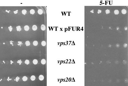
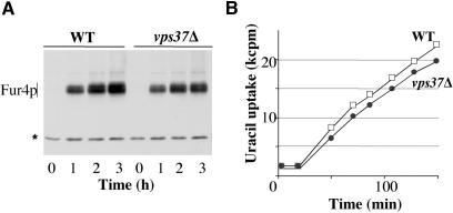
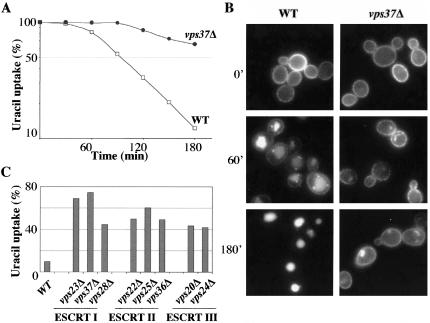
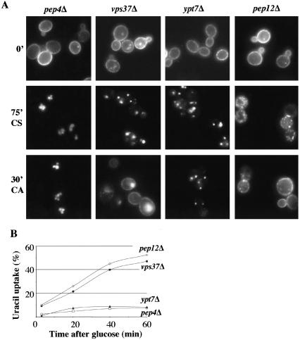
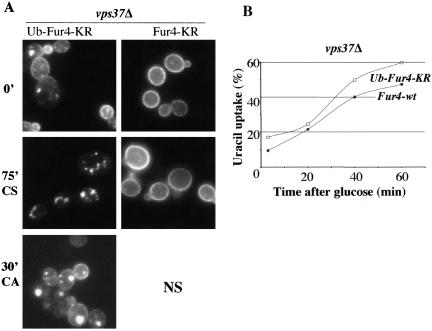
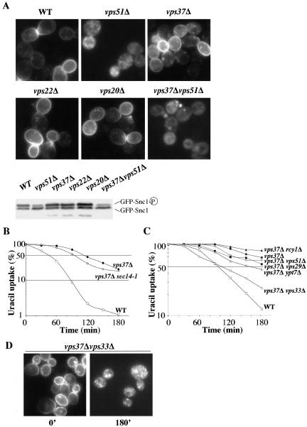
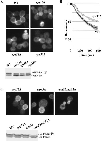
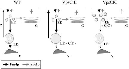
In Saccharomyces cerevisiae, deficiencies in the ESCRT machinery trigger the mistargeting of endocytic and biosynthetic ubiquitinated cargoes to the limiting membrane of the vacuole. Surprisingly, impairment of this machinery also leads to the accumulation of various receptors and transporters at the plasma membrane in both yeast and higher eukaryotes. Using the well-characterized yeast endocytic cargo uracil permease (Fur4p), we show here that the apparent stabilization of the permease at the plasma membrane in ESCRT mutants results from an efficient recycling of the protein. Whereas several proteins as well as internalized dyes are known to be recycled in yeast, little is known about the machinery and molecular mechanisms involved. The SNARE protein Snc1p is the only cargo for which the recycling pathway is well characterized. Unlike Snc1p, endocytosed Fur4p did not pass through the Golgi apparatus en route to the plasma membrane. Although ubiquitination of Fur4p is required for its internalization, deubiquitination is not required for its recycling. In an attempt to identify actors in this new recycling pathway, we found an unexpected phenotype associated with loss of function of the Vps class C complex: cells defective for this complex are impaired for recycling of Fur4p, Snc1p, and the lipophilic dye FM4-64. Genetic analyses indicated that these phenotypes were due to the functioning of the Vps class C complex in trafficking both to and from the late endosomal compartment.


Figures

Figure 1.
Figure 1.
ESCRT mutant cells are hypersensitive to 5-FU. BY4741 (WT), yJMG408 (vps37Δ, ESCRT I), yJMG475 (vps22Δ, ESCRT II), yJMG400 (vps20Δ, ESCRT III) cells transformed with empty vector and BY4741 (WT) transformed with Yep352fF (end-FUR4), allowing overproduction of Fur4p (x pFUR4), were tested for sensitivity to 5-FU. Exponentially growing cells were serially diluted and spotted onto SD plates without (-, left) or with 2.5 μM 5-FU (5-FU, right).
Figure 2.
Figure 2.
Fur4p secretion is not modified in ESCRT mutant cells. BY4741 (WT, white square) and yJMG408 (vps37Δ, black circle) cells transformed with p195GF (GAL-Fur4) were cultured in minimal media supplemented with raffinose until the early exponential growth phase. Fur4p synthesis was induced by adding galactose. (A) Crude extracts were prepared every hour after the addition of galactose, and Fur4p was detected by Western immunoblotting. The asterisk (*) indicates a nonspecific cross-reacting protein used as a loading control. (B) Uracil uptake was measured every 25 min over 150 min, results are expressed in kCPM.
Figure 3.
Figure 3.
Fur4p is stabilized at the plasma membrane of ESCRT mutant cells. The indicated strains transformed with pFL38gFP (GAL-Fur4-GFP) were cultured and Fur4-GFP synthesis was induced as described Figure 2. Glucose was added to block Fur4-GFP synthesis. The cells were incubated for 20 min, and rapamycin was then added to trigger endocytosis of the permease (t = 0). (A) Uracil uptake was measured every 30 min after the addition of rapamycin for 3 h in BY4741 (WT, white square) and yJMG408 (vps37Δ, black circle) cells. Results are expressed as a percentage of initial uptake at t = 0 on a semilog scale. (B) Cells were collected at the times indicated after rapamycin addition and visualized by fluorescence microscopy. (C) BY4741 (WT); yJMG401 (vps23Δ), yJMG408 (vps37Δ), and yJMG404 (vps28Δ) (ESCRT I); yJMG475 (vps22Δ), yJMG403 (vps25Δ), and yJMG407 (vps36Δ) (ESCRT II); yJMG400 (vps20Δ) and yJMG402 (vps24Δ) (ESCRT III) cells were treated as described above. Residual uracil uptake 3 h after rapamycin addition is plotted. Results are expressed as a percentage of initial uracil uptake at t = 0.
Figure 4.
Figure 4.
Fur4p is efficiently recycled to the plasma membrane of ESCRT mutant cells. yJMG503 (pep4Δ, white square), yJMG408 (vps37Δ, black circle), yJMG367 (ypt7Δ, black triangle), and yJMG498 (pep12Δ, white diamond) cells transformed with pFL38gFP (GAL-Fur4-GFP) were cultured at 24°C, and Fur4-GFP synthesis was induced as described in Figure 3. At t = 0, cells were subjected to carbon starvation to trigger endocytosis of the permease, and glucose was added 75 min later. (A) Cells were visualized by fluorescence microscopy at t = 0 (0′), after 75 min of carbon starvation (75′CS) and 30 min after the addition of carbon (30′CA). Note that Fur4-GFP was efficiently internalized in all the strains and the protein was rapidly targeted back to the plasma membrane in vps37Δ and pep12Δ cells after the addition of glucose. (B) Uracil uptake was measured every 20 min after the addition of glucose. Results are expressed as a percentage of the initial uracil uptake measured immediately before carbon starvation.
Figure 5.
Figure 5.
Deubiquitination of Fur4p is not required for its recycling. yJMG408 (vps37Δ) cells transformed either with pFL38-KR-gFP (GAL-Fur4KR-GFP) or with pFL38gFPUb-KR (GAL-Ub-Fur4KR-GFP) were treated as described in Figure 4. (A) Cells were visualized by fluorescence microscopy at t = 0 (0′), after 75 min of carbon starvation (75′CS) and 30 min after the addition of carbon (30′CA) (left). Note that because Fur4-KR remained stable at the plasma membrane upon carbon starvation, the recycling of this mutant form cannot be followed (NS). (B) Uracil uptake was measured every 20 min after glucose addition in yJMG408 (vps37Δ) cells transformed with pFL38gFP-Ub-KR (GAL-Ub-Fur4KR-GFP, white square). To facilitate comparison, the curve from Figure 4B corresponding to yJMG408 (vps37Δ) transformed with pFL38gFP (GAL-Fur4-GFP, black circle) is replotted here. Results are expressed as a percentage of the initial uracil uptake measured immediately before carbon starvation.
Figure 6.
Figure 6.
Fur4p and Snc1p follow different recycling roads. (A) BY4741 (WT), yJMG453 (vps51Δ, VFT), yJMG408 (vps37Δ, ESCRT I), yJMG475 (vps22Δ, ESCRT II), yJMG400 (vps20Δ, ESCRT III), and yJMG531 (vps37Δvps51Δ, ESCRT I, VFT) cells were transformed with pJMG118 (tpi-GFP-Snc1) and cultured until the early exponential growth phase. The subcellular distribution of GFPSnc1p was monitored by fluorescence microscopy (top). Crude extracts were prepared and the phosphorylation state of GFP-Snc1p was checked by Western immunoblotting in the indicated strains (bottom). (B) BY4741 (WT, white square), yJMG524 (vps37Δsec14-1, white diamond), and yJMG408 (vps37Δ, black circle) transformed with pFL38gFP (GAL-Fur4-GFP) were grown at 24°C and preshifted at 37° 15 min before addition of rapamycin. Uracil uptake was measured every 30 min after the addition of rapamycin over a 3-h period; results are expressed as a percentage of the initial uptake at t = 0 in a semilog scale. (C) yJMG528 (vps37Δrcy1Δ, black triangle), yJMG530 (vps37Δypt7Δ, white triangle), (yJMG531 (vps37Δvps51Δ, white diamond), yJMG529 (vps37Δvps29Δ, black square) and yJMG523 (vps37Δvps33Δ, white circle) cells were transformed, cultured, induced, and treated with rapamycin as described in Figure 3. The curves from Figure 3A corresponding to BY4741 (WT, white square) and yJMG408 (vps37Δ, black circle) transformed with pFL38gFP (GAL-Fur4-GFP) are replotted here. (D) yJMG523 (vps37Δvps33Δ) cells were collected at the time indicated after the addition of rapamycin and visualized by fluorescence microscopy. Note that Fur4-GFP accumulated in intracellular dots at t = 180′ (compare with Figure 3B).
Figure 7.
Figure 7.
Multiple recycling defects in Vps class C mutant cells. (A) BY4741 (WT) and yJMG473 (vps16Δ), yJMG474 (vps18Δ), yJMG476 (vps33Δ) cells defective for the VpsClC complex, transformed with pJMG118 (tpi-GFP-Snc1) were cultured and analyzed for GFP-Snc1p recycling as described in Figure 6A. Note that GFP-Snc1p was mainly intracellular (top) and underphosphorylated (bottom) in all strains defective for the VpsClC complex tested. (B) BY4741 (WT) and yJMG476 (vps33Δ) cells were analyzed for their ability to recycle the fluorescent membrane dye FM4-64, as described in Materials and Methods. Fluorescence was measured in triplicate for each strain, every 6 s for 10 min on a spectrofluorimeter. Results are expressed as a percentage of the initial fluorescence ± SD. (C) yJMG498 (pep12Δ), yJMG506 (vam3Δ), and yJMG526 (vam3Δpep12Δ) cells transformed with pJMG118 (tpi-GFP-Snc1, URA3, and CEN) were cultured and analyzed for GFP-Snc1p recycling as described in Figure 6A. Note that GFP-Snc1p displayed a similar distribution and phosphorylation defect in vam3Δpep12Δ cells and VpsClC-deficient cells (see A).
Figure 8.
Figure 8.
Working model. Fur4p (black arrow) and Snc1p (gray arrow) recycle via different pathways. The thickness of the arrows reflects the intensity of the traffic. Left, in wt cells: Fur4p follows the endocytic pathway through EE, LE, and is degraded in the vacuole (V), with a small amount possibly recycling (?). Snc1p constitutively cycles between EE, the Golgi apparatus (G) and the plasma membrane (PM). Middle, in VpsClE mutant cells: after endocytosis, Fur4p recycles efficiently from the ClE compartment to the PM. Snc1p recycling is not affected. Right, in VpsClC mutant cells: after endocytosis, Fur4p and Snc1p are blocked in a modified early endosomal compartment (EE “ClC”) from which they cannot recycle to the plasma membrane. See text for details.

Similar articles

Cited by

References

    1. Amerik, A.Y., Nowak, J., Swaminathan, S., and Hochstrasser, M. (2000). The Doa4 deubiquitinating enzyme is functionally linked to the vacuolar protein sorting and endocytic pathways. Mol. Biol. Cell 11, 3365-3380. - PMC - PubMed
    1. Andrews, N. (2002). Lysosomes and the plasma membrane: trypanosomes reveal a secret relationship. J. Cell Biol. 158, 389-394. - PMC - PubMed
    1. Babst, M., Odorizzi, G., Estepa, E.J., and Emr, S.D. (2000). Mammalian tumor susceptibility gene 101 (TSG101) and the yeast homologue, Vps23p, both function in late endosomal trafficking. Traffic 1, 248-258. - PubMed
    1. Bensen, E.S., Yeung, B.G., and Payne, G.S. (2001). Ric1p and the Ypt6p GTPase function in a common pathway required for localization of trans-Golgi network membrane proteins. Mol. Biol. Cell 12, 13-26. - PMC - PubMed
    1. Bilodeau, P.S., Urbanowski, J.L., Winistorfer, S.C., and Piper, R.C. (2002). The Vps27p Hse1p complex binds ubiquitin and mediates endosomal protein sorting. Nat. Cell Biol. 4, 534-539. - PubMed

Publication types

MeSH terms