

PDBSiteScan: a program for searching for active, binding and posttranslational modification sites in the 3D structures of proteins
- PMID: 15215447
- PMCID: PMC441577
- DOI: 10.1093/nar/gkh439
PDBSiteScan: a program for searching for active, binding and posttranslational modification sites in the 3D structures of proteins
Abstract
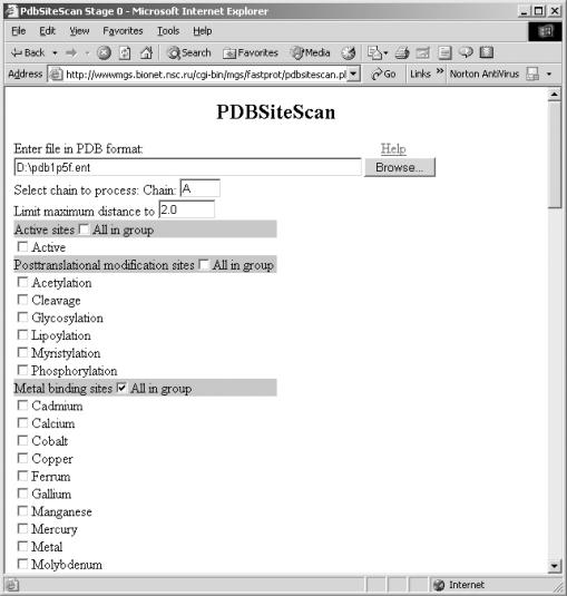
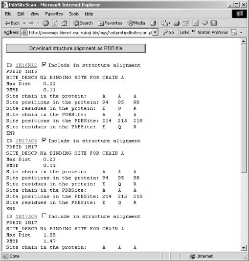
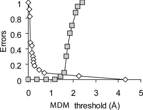
PDBSiteScan is a web-accessible program designed for searching three-dimensional (3D) protein fragments similar in structure to known active, binding and posttranslational modification sites. A collection of known sites we designated as PDBSite was set up by automated processing of the PDB database using the data on site localization in the SITE field. Additionally, protein-protein interaction sites were generated by analysis of atom coordinates in heterocomplexes. The total number of collected sites was more than 8100; they were assigned to more than 80 functional groups. PDBSiteScan provides automated search of the 3D protein fragments whose maximum distance mismatch (MDM) between N, Calpha and C atoms in a fragment and a functional site is not larger than the MDM threshold defined by the user. PDBSiteScan requires perfect matching of amino acids. PDBSiteScan enables recognition of functional sites in tertiary structures of proteins and allows proteins with functional information to be annotated. The program PDBSiteScan is available at http://wwwmgs.bionet.nsc.ru/mgs/systems/fastprot/pdbsitescan.html.
Figures
References
-
- Fetrow J.S. and Skolnick,J. (1998) Method for prediction of protein function from sequence using the sequence-to-structure-to-function paradigm with application to glutaredoxins/thioredoxins and T1 ribonucleases. J. Mol. Biol., 281, 949–68. - PubMed
-
- Todd A.E., Orengo,C.A. and Thornton,J.M. (1999) DOMPLOT: a program to generate schematic diagrams of the structural domain organization within proteins, annotated by ligand contacts. Protein Eng., 12, 375–379. - PubMed
-
- Holm L. and Sander,C. (1997) An evolutionary treasure: unification of a broad set of amidohydrolases related to urease. Proteins, 28, 72–82. - PubMed
MeSH terms
Substances
Associated data
- Actions
LinkOut - more resources
Full Text Sources
Miscellaneous
