. 2004 Jul;13(7):1735-49.
doi: 10.1110/ps.04651004.

Determinants of quaternary association in legume lectins

Affiliations

Determinants of quaternary association in legume lectins

K V Brinda et al. Protein Sci. 2004 Jul.

Abstract

It is well known that the sequence of amino acids in proteins code for its tertiary structure. It is also known that there exists a relationship between sequence and the quaternary structure of proteins. The question addressed here is whether the nature of quaternary association can be predicted from the sequence, similar to the three-dimensional structure prediction from the sequence. The class of proteins called legume lectins is an interesting model system to investigate this problem, because they have very high sequence and tertiary structure homology, with diverse forms of quaternary association. Hence, we have used legume lectins as a probe in this paper to (1) gain novel insights about the relationship between sequence and quaternary structure; (2) identify the sequence motifs that are characteristic of a given type of quaternary association; and (3) predict the quaternary association from the sequence motif.


Figures
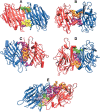
Figure 1.
Figure 1.
Examples of the five major types of interfaces in legume lectins. A–E represents the three-dimensional structure of the legume lectins belonging to these five types of dimeric interfaces. The monomer chains are represented in the form of cartoon diagrams in blue and in red color, respectively. Although the tertiary structures are similar, in all the five cases, their quaternary associations are different as seen in the figure. The interface cluster-forming residues are represented as van der Waals’ spheres. Each cluster is colored differently to differentiate them in the three-dimensional space. (A) Canonical—type II (1fatAB at 6% cutoff); (B) ECorL-type—X3 (1axy at 5% cutoff); (C) GS4-type—X4 (1gsl at 6% cutoff); (D) DB58-type—X1 (1qnwAC at 4% cutoff); (E) Noncanonical interface of ConA-type—X2 (1dglAC at 4% cutoff).
Figure 2.
Figure 2.
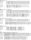
Multiple sequence alignments of different legume lectin interface types. (A) Canonical (II); (B) ECorL-type (X3); (C) GS4-type (X4); (D) DB58-type (X1); (E) Noncanonical interface of ConA-type (X2). For want of space, only some examples in each case and the important regions involved are shown in the figure, that is, N terminus and C terminus, region 50–80, and region 150–200. The alignment also includes an example of a lectin with unknown structure (LUS), wherever available. The residue number of each protein in the aligned region is given at the end of each line. The complete conservation, partial conservation, and conservative mutations that occur in the sequence alignment are shown as “*”, “.”, and “:”, respectively. The residues, which are either completely or partially conserved or conservatively mutated in sequence and are also present in the interface clusters of the legume lectins belonging to a particular category, are shown in bold and underlined.
Figure 3.
Figure 3.
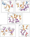
Interface cluster-forming residues in the five major types of legume lectin interfaces. (A) Canonical—type II (1fatAB at 6% cutoff); (B) ECorL-type—X3 (1axy at 5% cutoff); (C) GS4-type—X4 (1gsl at 6% cutoff); (D) DB58-type—X1 (1qnwAC at 4% cutoff); (E) Noncanonical interface of ConA-type—X2 (1dglAC at 4% cutoff). The representations are in a ball-and-stick model in the entire figure. Due to constraint of space, only one interface cluster is shown in each example, although there may actually be more than one interface cluster. The coloring scheme is as follows: blue, residues from chain A that are not conserved in the interface clusters of other proteins of the same interface type; red, residues from chain B/C/D that are not conserved in the interface clusters of other proteins of the same interface type; gold, residues from chain A that are conserved (in sequence and structure by total or partial conservation or conservative mutation) in the interface clusters of other proteins of the same interface type; magenta, residues from chain B/C/D that are conserved (in sequence and structure by total or partial conservation or conservative mutation) in the interface clusters of other proteins of the same interface type. The residue number, name and chain are indicated in the figure. The position of the vector components of the top eigenvalues corresponding to these interface cluster-forming residues is also indicated within parentheses. We see that the residues colored in gold and in magenta have higher vector component magnitude than the ones colored in blue and in red, indicating that the residues that are important for the stability of the cluster and the interface (indicated by high vector component magnitude) and the ones that are conserved in the interface clusters (shown in gold and magenta) correlate very well.

Similar articles

Cited by

References

    1. Banerjee, R., Mande, S.C., Ganesh, V., Das, K., Dhanaraj, V., Mahanta, S.K., Suguna, K., Surolia, A., and Vijayan, M. 1994. Crystal structure of peanut lectin, a protein with an unusual quaternary structure. Proc. Natl. Acad. Sci. 91 227–231. - PMC - PubMed
    1. Berman, H.M., Westbrook, J., Feng, Z., Gilliland, G., Bhat, T.N., Weissig, H., Shindyalov, I.N., and Bourne, P.E. 2000. The Protein Data Bank. Nucleic Acids Res. 28 235–242. - PMC - PubMed
    1. Boeckmann, B., Bairoch, A., Apweiler, R., Blatter, M.-C., Estreicher, A., Gasteiger, E., Martin, M.J., Michoud, K., O’Donovan, C., Phan, I., et al. 2003. The SWISS-PROT protein knowledgebase and its supplement TrEMBL in 2003. Nucleic Acids Res. 31 365–370. - PMC - PubMed
    1. Brinda, K.V., Kannan, N., and Vishveshwara, S. 2002. Analysis of homodimeric protein interfaces by graph-spectral methods. Protein Eng. 4 265–277. - PubMed
    1. Buts, L., Dao-Thi, M.H., Loris, R., Wyns, L., Etzler, M., and Hamelryck, T. 2001. Weak protein–protein interactions in lectins: The crystal structure of a vegetative lectin from the legume Dolichos biflorus. J. Mol. Biol. 309 193–201. - PubMed

Publication types

Associated data

LinkOut - more resources