. 2004 Jul;13(7):1802-10.
doi: 10.1110/ps.04679104.

Removal of N-terminal methionine from recombinant proteins by engineered E. coli methionine aminopeptidase

Affiliations

Removal of N-terminal methionine from recombinant proteins by engineered E. coli methionine aminopeptidase

You-Di Liao et al. Protein Sci. 2004 Jul.

Abstract

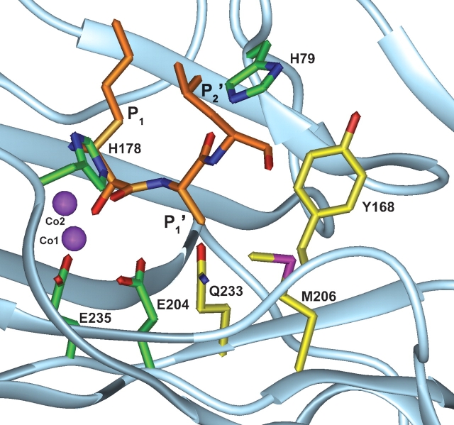
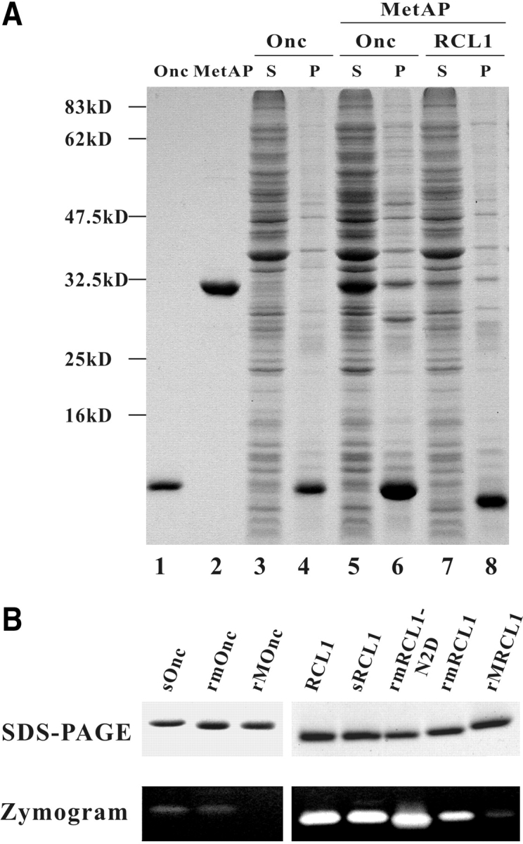
The removal of N-terminal translation initiator Met by methionine aminopeptidase (MetAP) is often crucial for the function and stability of proteins. On the basis of crystal structure and sequence alignment of MetAPs, we have engineered Escherichia coli MetAP by the mutation of three residues, Y168G, M206T, Q233G, in the substrate-binding pocket. Our engineered MetAPs are able to remove the Met from bulky or acidic penultimate residues, such as Met, His, Asp, Asn, Glu, Gln, Leu, Ile, Tyr, and Trp, as well as from small residues. The penultimate residue, the second residue after Met, was further removed if the antepenultimate residue, the third residue after Met, was small. By the coexpression of engineered MetAP in E. coli through the same or a separate vector, we have successfully produced recombinant proteins possessing an innate N terminus, such as onconase, an antitumor ribonuclease from the frog Rana pipiens. The N-terminal pyroglutamate of recombinant onconase is critical for its structural integrity, catalytic activity, and cyto-toxicity. On the basis of N-terminal sequence information in the protein database, 85%-90% of recombinant proteins should be produced in authentic form by our engineered MetAPs.


Figures

Figure 1.
Figure 1.
Diagram of the substrate-binding pocket of E. coli MetAP. Part of E. coli MetAP (in light blue), two cobalt ions (in purple), and bestatin-based inhibitor (P1, P1′, and P2′ in orange) complex is adapted from PDB database (3MAT). The residues His 79, His 178, Glu 204, and Glu 235, involved in catalytic activity, and the residues Tyr 168, Met 206, and Gln 233, involved in substrate recognition, are colored green and yellow, respectively.
Figure 2.
Figure 2.
Expression and analyses of MetAP and target ribonucleases. (A) Coexpression of MetAP and target ribonucleases. The protein component of supernatant and insoluble pellet of 30-μL transformed E. coli BL21(DE3) culture was analyzed by 13.3% SDS-PAGE and Coomassie blue staining. (Lane 1) Two micrograms onconase; (lane 2) three micrograms MetAP; (lanes 3,4) supernatant and pellet of E. coli expressing onconase only; (lanes 5,6) supernatant and pellet of E. coli coexpressing engineered MetAP and onconase; (lanes 7,8) supernatant and pellet of E. coli coexpressing engineered MetAP and RC-RNaseL1. S and P represent the supernatant and insoluble pellet of E. coli lysate, respectively. (B) Analyses of recombinant ribonucleases. (Top panel) Two micrograms of purified ribonuclease was separated by 13.3% SDS-PAGE and stained by Coomassie blue. (Bottom panel) Ten nanograms onconase and 1 ng RC-RNaseL1 were separated by RNA-casting SDS-PAGE and stained by Toluidine blue O. sOnc and sRCL1 represent secretory onconase and RC-RNase L1, respectively, which were purified from culture medium; rmOnc, rmRCL1, and rmRCL1-N2D represent Met-tagged onconase, RC-RNase L1, and RC-RNase L1-N2D, respectively, which are treated with MetAP-*TG in vivo; rMOnc and rMRCL1 represent Met-tagged onconase and RC-RNase L1, respectively, without MetAP treatment; RCL1 represents native RC-RNase L1 purified from bullfrog liver.

Similar articles

Cited by

References

    1. Abe, A., Saeki, K., Yasunaga, T., and Wakabayashi, T. 2000. Acetylation at the N-terminus of actin strengthens weak interaction between actin and myosin. Biochem. Biophys. Res. Commun. 268 14–19. - PubMed
    1. Adachi, K., Yamaguchi, T., Yang, Y., Konitzer, P.T., Pang, J., Reddy, K.S., Ivanova, M., Ferrone, F., and Surrey, S. 2000. Expression of functional soluble human β-globin chains of hemoglobin in bacteria. Protein Expr. Purif. 20 37–44. - PubMed
    1. Belagaje, R.M., Reams, S.G., Ly, S.C., and Prouty, W.F. 1997. Increased production of low molecular weight recombinant proteins in Escherichia coli. Protein Sci. 6 1953–1962. - PMC - PubMed
    1. Ben-Bassat, A., Bauer, K., Chang, S.Y., Myambo, K., Boosman, A., and Chang, S. 1987. Processing of the initiation methionine from proteins: Properties of the Escherichia coli methionine aminopeptidase and its gene structure. J. Bacteriol. 169 751–757. - PMC - PubMed
    1. Boix, E., Wu, Y., Vasandani, V.M., Saxena, S.K., Ardelt, W., Ladner, J., and Youle, R.J. 1996. Role of the N terminus in RNase A homologues: Differences in catalytic activity, ribonuclease inhibitor interaction and cytotoxicity. J. Mol. Biol. 257 992–1007. - PubMed

MeSH terms

LinkOut - more resources