Comparative Study
. 2004 Aug 15;559(Pt 1):301-13.
doi: 10.1113/jphysiol.2004.064998. Epub 2004 Jun 24.

Mechanisms of deep brain stimulation: an intracellular study in rat thalamus

Affiliations
Comparative Study

Mechanisms of deep brain stimulation: an intracellular study in rat thalamus

Trent Anderson et al. J Physiol. .

Abstract

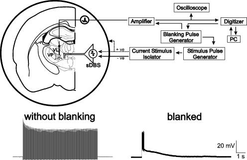
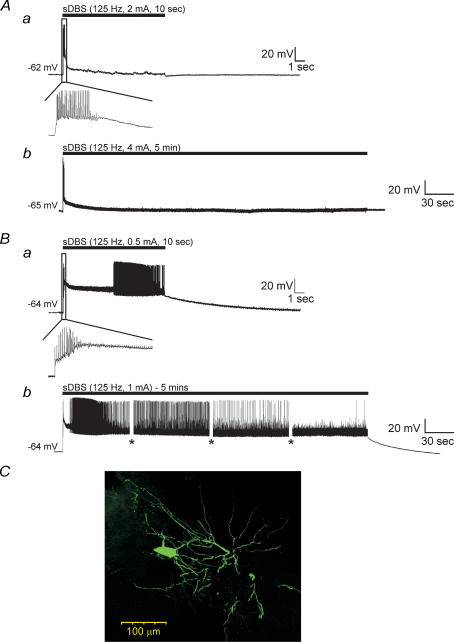
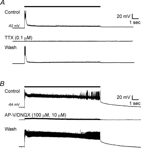
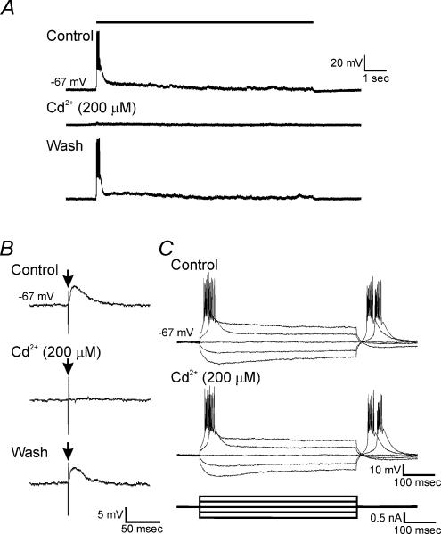
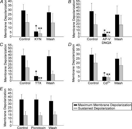
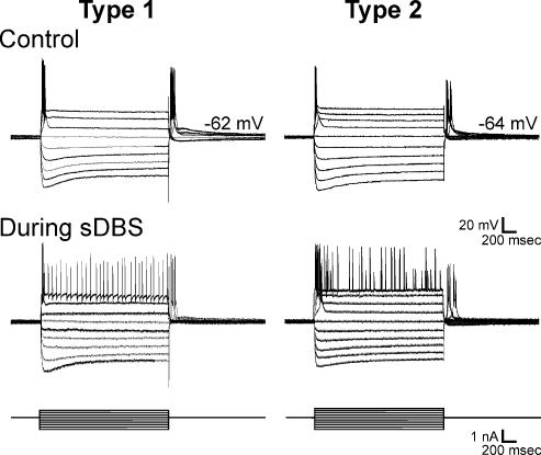
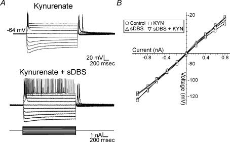
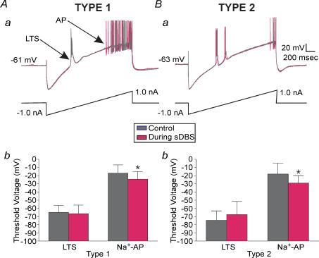
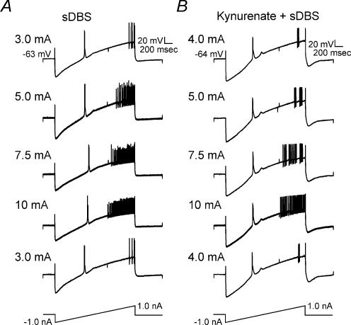
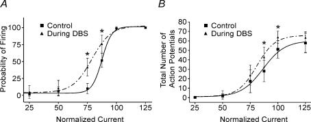
High-frequency deep brain stimulation (DBS) in the thalamus alleviates most kinds of tremor, yet its mechanism of action is unknown. Studies in subthalamic nucleus and other brain sites have emphasized non-synaptic factors. To explore the mechanism underlying thalamic DBS, we simulated DBS in vitro by applying high-frequency (125 Hz) electrical stimulation directly into the sensorimotor thalamus of adult rat brain slices. Intracellular recordings revealed two distinct types of membrane responses, both of which were initiated with a depolarization and rapid spike firing. However, type 1 responses repolarized quickly and returned to quiescent baseline during simulated DBS whereas type 2 responses maintained the level of membrane depolarization, with or without spike firing. Individual thalamic neurones exhibited either type 1 or type 2 response but not both. In all neurones tested, simulated DBS-evoked membrane depolarization was reversibly eliminated by tetrodotoxin, glutamate receptor antagonists, and the Ca(2+) channel antagonist Cd(2+). Simulated DBS also increased the excitability of thalamic cells in the presence of glutamate receptor blockade, although this non-synaptic effect induced no spontaneous firing such as that found in subthalamic nucleus neurones. Our data suggest that high-frequency stimulation when applied in the ventral thalamus can rapidly disrupt local synaptic function and neuronal firing thereby leading to a 'functional deafferentation' and/or 'functional inactivation'. These mechanisms, driven primarily by synaptic activation, help to explain the paradox that lesions, muscimol and DBS in thalamus all effectively stop tremor.


Figures

Figure 1
Figure 1. Experimental methods
Top: schematic representation of sharp intracellular recording set-up in thalamic rat brain slice. Note the two methods of stimulation using either the intranuclear bipolar electrode or the surrounding ‘monopolar-ring’ configuration. Recording and stimulation sites were in ventral-lateral (VL) or ventral-posterior (VP) nuclei. Bottom: intracellular recordings from the same cell with, and without the blanking operation activated.
Figure 2
Figure 2. sDBS evoked two distinct types of membrane responses in ventral thalamic neurones
A, type 1 responses had a large initial depolarization declining toward a smaller but sustained level of depolarization in response to 10 s (a) or 5 min (b) of sDBS. The black bar indicates stimulus onset and duration. B, type 2 responses have a large initial depolarization, which persisted over 10 s (a) or 5 min (b) and led to varied spike activity. The insets show expanded initial responses during the 10 s sDBS train. The amplitude of action potentials in this and following figures were partially reduced due to digitization and some were also truncated or flattened by ‘blanking pulses’. Gaps in the recording (shown as *) indicate times at which current pulse protocols were run during the 5 min sDBS train. C, morphology of a representative ventral thalamic neurone filled with neurobiotin. Note the extensive dendritic tree with numerous distal arborizations.
Figure 3
Figure 3. Pharmacological blockade of membrane responses induced by sDBS
Glutamate receptor (kynurenate, or AP-V–DNQX) or Na+ channel (TTX) blockade were equally effective in preventing type 1 and 2 responses. A, type 1 response before, during and after washout of TTX. B, type 2 response to sDBS before, during and after washout of bath application of AP-V–DNQX. Note that glutamatergic blockade also eliminated action potentials induced by sDBS.
Figure 4
Figure 4. Cd2+ selectively blocked sDBS responses
A, membrane responses to sDBS before, during bath application of Cd2+, and after washout. B, representative trace of single shock-evoked EPSPs before, during bath application of 200 μm Cd2+, and after washout. Vertical excursion, indicated by arrow (↓), is stimulus artifact. C, current injection pulses recorded before and during bath application of 200 μm Cd2+. Note that the T-type Ca2+ channel-dependent LTS remains unchanged in the presence of 200 μm Cd2+. All recordings were obtained from the same cell.
Figure 5
Figure 5. Summarized pharmacological data showing blockade of sDBS-induced maximal and sustained depolarization
Depolarization is almost eliminated in the presence of: kynurenate (2 mm; n = 10; A), AP-V–DNQX (100 μm–10 μm; n = 6; B), TTX (0.1 μm; n = 7; C), Cd2+ (200 μm; n = 6; D). Picrotoxin (50 μm; n = 4; E) does not affect membrane depolarization. Because both type 1 and 2 responding cells had similar suppression of depolarization, all data are presented together. Responses under pharmacological treatment that were statistically different from their respective control and washout values are indicated (* initial depolarization, ** mean sustained depolarization, P < 0.05).
Figure 6
Figure 6. Effects of transmembrane current pulses
A, intracellular transmembrane current pulses (1 s) were applied to neurones previously identified as having a type 1 (n = 8) or type 2 (n = 3) membrane response to sDBS. Identical current pulses were re-applied during sDBS. Manual current was injected to offset the depolarization induced by sDBS. Note the similarity to transmembrane current pulse responses in both type 1 and 2 responding neurones, indicative of a common cell type.
Figure 7
Figure 7. Effects of sDBS on membrane conductance
A, step current injections were used to obtain a I–V curve both during control sDBS or in the presence of kynurenate. Similar results were obtained for both type 1 and type 2 sDBS membrane responses. A representative trace from a type 2 response is displayed. B, no significant change in steady-state conductance during control (○), sDBS (Δ), kynurenate (□), or sDBS + kynurenate (∇). Each data point is derived from a minimum of 5 cells.
Figure 8
Figure 8. Decrease in firing threshold during sDBS
Overlayed recordings of membrane responses to ramp current injections (2 s, −1.0 to 1.0 nA) in type 1 (Aa) and type 2 (Ba) cells in control and during sDBS conditions. A significant decrease in the firing threshold for Na+ spikes (P < 0.05) during sDBS but not in the low threshold Ca2+ spikes (LTS) was found with both response types. The summarized data are shown in Ab (type 1; n = 7) and Bb (type 2; n = 4).
Figure 9
Figure 9. sDBS-induced increase in firing rate is dependent on stimulation strength, but independent of glutamate receptor activation
A, sDBS was applied at different intensities in the same type 1 neurone while spike firing rate was evaluated using ramp current injection. Increasing sDBS current amplitude significantly increased the firing rate. Note that comparable responses to ramp current injection were elicited in both A and B when initial sDBS current intensities were retested after high currents (up to 10 mA) were used. This indicates that no adverse membrane effect resulted from high sDBS current levels. B, the same firing rate tests as inA was applied in another neurone where glutamatergic transmission was blocked with 2 mm kynurenate.
Figure 10
Figure 10
sDBS-induced increase in firing probability and frequency in a type 1 response A train of 30 intracellular current pulses (3.3 Hz, 100 ms width) with varying intensities (50–1000 pA) were applied before (▪) and during sDBS (▴). The firing probability and rate for each current level were calculated. Current was normalized to the intensity level that produced 100% probability of firing (see Results). The total number of action potentials per series of 30 intracellular pulses was examined. The presence of single action potentials prevented the use of interspike interval as a measure of frequency. Summarized data (n = 3 each) showing a significant increase in firing probability (A) and frequency (B) during sDBS are reported (P < 0.05).

Similar articles

Cited by

References

    1. Anderson T, Hu B, Pittman Q, Kiss ZHT. Synaptic activation by deep brain stimulation: An intracellular study in rat thalamus. Soc Neurosci Abstract. 2003b 185.4. - PMC - PubMed
    1. Anderson ME, Postupna N, Ruffo M. Effects of high-frequency stimulation in the internal globus pallidus on the activity of thalamic neurons in the awake monkey. J Neurophysiol. 2003a;89:1150–1160. - PubMed
    1. Benabid AL, Pollak P, Gao DM, Hoffmann D, Limousin P, Gay E, Payen I, Benazzouz A. Chronic electrical stimulation of the ventralis intermedius nucleus of the thalamus as a treatment of movement disorder. J Neurosurg. 1996;84:203–214. - PubMed
    1. Bergman H, Deuschl G. Pathophysiology of Parkinson's disease: from clinical neurology to basic neuroscience and back. Mov Disord. 2002;17(Suppl. 3):S28–S40. - PubMed
    1. Beurrier C, Bioulac B, Audin J, Hammond C. High-frequency stimulation produces a transient blockade of voltage-gated currents in subthalamic neurons. J Neurophysiol. 2001;85:1351–1356. - PubMed

Publication types

LinkOut - more resources