Comparative Study
. 2004 Aug 15;559(Pt 1):259-69.
doi: 10.1113/jphysiol.2004.063016. Epub 2004 Jun 24.

Properties of unitary potentials generated by intramuscular interstitial cells of Cajal in the murine and guinea-pig gastric fundus

Affiliations
Comparative Study

Properties of unitary potentials generated by intramuscular interstitial cells of Cajal in the murine and guinea-pig gastric fundus

E A H Beckett et al. J Physiol. .

Abstract

Intracellular recordings were made from isolated bundles of the circular muscle layer of mouse and guinea-pig gastric fundus. These preparations displayed an ongoing discharge of membrane noise (unitary potentials), similar to that recorded from similar preparations made from the circular layer of the antrum. Bundles of muscle from the fundus of W/W(V) mice, which lack intramuscular interstitial cells of Cajal (ICC(IM)) lacked the discharge of membrane noise observed in wild-type tissues. When the membrane potential was changed by passing depolarizing or hyperpolarizing current pulses, the discharge of membrane noise was little changed. The membrane noise was unaffected by adding chloride channel blockers; however, agents which buffered the internal concentration of calcium ions reduced the discharge of membrane noise. Treatment of tissues with CCCP, which interferes with the uptake of calcium ions by mitochondria, also reduced the membrane noise and caused membrane hyperpolarization. Similar observations were made on bundles of tissue isolated from the circular layer of the guinea pig antrum. Together the observations indicate that membrane noise is generated by a pathway located in ICC(IM). The properties of this pathway appear to vary dramatically within a given organ. The lack of voltage sensitivity of the discharge of membrane noise in the fundus provides a possible explanation for the lack of rhythmic electrical activity in this region of the stomach.


Figures
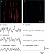
Figure 1
Figure 1. Distribution of kit positive cells in bundles of circular muscle isolated from the fundus of C57BL/6 and W/WV mice and the discharge of membrane noise
A and E, the distribution of Kit-immunopositive ICCIM in bundles of circular muscle dissected from the fundus of a C57BL/6 mouse (A) and a W/WV mouse (E). Elongated, spindle-shaped ICCIM running parallel to the circular muscle fibres were readily detected in preparations obtained from C57BL/6 mice (A) but not in W/WV mice (E). A and E are composites of 9 μm × 0.8 μm optical sections through the circular muscle bundles. The white dotted lines denote the border of the muscle bundles in both A and E. The 50 μm scale bar in E is for both panels A and E. Samples of membrane noise recorded from a circular muscle bundle isolated from the fundus of a C57BL/6 mouse in control conditions, in the presence of nifedipine (1 μm) and in the presence of both nifedipine and apamin (0.2 μm) are shown in B, C and D, respectively. Recordings from a bundle isolated from W/WV fundus in control conditions, in the presence of nifedipine (1 μm) and in the presence of both nifedipine and apamin (0.2 μm) are shown in F, G and H, respectively. The calibration bars shown in panel H also refer to panels BD and FG.
Figure 2
Figure 2. Power spectral density curves calculated from recordings made from wild-type and W/WV fundus muscle bundles
Samples of membrane noise recorded from a circular muscle bundle isolated from the fundus of a C57BL/6 mouse (A) and a W/WV mouse (C) in the presence of nifedipine and apamin are shown in A and C, respectively. B and D, power spectral density curves derived from wild-type (A) and W/WV (C) recordings, respectively.
Figure 3
Figure 3. Electrical properties of a bundle of circular muscle isolated from a C57BL/6 wild-type fundus
A, averaged electrotonic potentials recorded from a wild-type circular muscle bundle produced by injecting hyperpolarizing currents. Break of membrane hyperpolarization failed to initiate regenerative potentials. B, altering the membrane potential over a range of potentials using depolarizing and hyperpolarizing current pulses also failed to elicit regenerative potentials.
Figure 4
Figure 4. Effect of changing the membrane potential on discharge of membrane noise in a bundle of circular muscle isolated from wild-type fundus
A, an averaged trace of 10 electrotonic potentials produced by injecting 1 nA of hyperpolarizing current into a circular muscle bundle. Three sample traces of individual electrotonic potentials are shown in B. Note that hyperpolarizing the bundles by over 20 mV failed to abolish the discharge of membrane noise. Hyperpolarized (a) and baseline regions of these traces (b) were used to construct the power spectral density curves shown in C and D (○, hyperpolarized region, a in panel C; •, baseline region, b in panel D). Note that hyperpolarization did not change the shape of the power spectral density curve but the power function was increased by approximately 100%.
Figure 5
Figure 5. Lack of effect of chloride channel blocker on membrane noise recorded from wild-type circular muscle bundle
Three traces of membrane noise recorded from a circular muscle bundle in control conditions (nifedipine; 1 μm) are shown in A and power spectra in B. The chloride channel blocker DIDS (100 μm) did not alter the discharge of membrane noise, demonstrated by representative traces (C) and the calculated power spectra (D).
Figure 6
Figure 6. Effect of buffering [Ca2+]i on discharge of membrane noise in a bundle of circular muscle isolated from the mouse fundus
A and B, membrane noise discharge recorded from a wild-type circular muscle bundle in control conditions and the calculated power spectra, respectively. BAPTA-AM (20 μm) dramatically reduced the frequency of membrane noise discharge until individual unitary potentials could be resolved (C). Power spectra curves calculated from BAPTA-AM traces demonstrated a substantial decrease in power (D). The power function had to be reduced by 50% of control to obtain an adequate fit in BAPTA-AM.
Figure 7
Figure 7. Effect of inhibiting Ca2+ uptake by mitochondria on discharge of membrane noise in a bundle of circular muscle isolated from the mouse fundus
Membrane noise recorded from a wild-type circular muscle bundle in control conditions and the corresponding power spectra curve are shown in A and B, respectively. C, the mitochondrial inhibitor CCCP (10 μm) abolished membrane noise activity. D, power spectra curve calculated from the traces recorded in the presence of CCCP.
Figure 8
Figure 8. Similarities between properties of membrane noise recorded from a circular muscle bundle isolated from guinea pig fundus and that recorded from a mouse circular muscle bundle
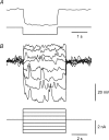
A, current injection pulses were used to vary the membrane potential of a circular muscle bundle of guinea pig fundus over a wide range of potentials (from 15 mV more positive than resting membrane potential to 40 mV more negative than resting membrane potential). Neither membrane depolarization, nor the break after 5 s of hyperpolarization initiated a regenerative potential. B, power spectral density curves calculated from membrane noise recorded before (•) and after (○) the addition of 9-AC could be fitted by the same function. C, power spectral density curves calculated from membrane noise recorded in control conditions (•) and in the presence of 30 μm BAPTA-AM (○). After 15 min in BAPTA-AM membrane noise was effectively abolished.

References

    1. Beckett EA, Horiguchi K, Khoyi M, Sanders KM, Ward SM. Loss of enteric motor neurotransmission in the gastric fundus of Sl/Sl(d) mice. J Physiol. 2002;543:871–887. - PMC - PubMed
    1. Beckett EA, McGeough CA, Sanders KM, Ward SM. Pacing of interstitial cells of Cajal in the murine gastric antrum: neurally mediated and direct stimulation. J Physiol. 2003;553:545–559. - PMC - PubMed
    1. Burns AJ, Herbert TM, Ward SM, Sanders KM. Interstitial cells of Cajal in the guinea-pig gastrointestinal tract as revealed by c-kit immunohistochemistry. Cell Tissue Res. 1997;290:11–20. - PubMed
    1. Burns AJ, Lomax AE, Torihashi S, Sanders KM, Ward SM. Interstitial cells of Cajal mediate inhibitory neurotransmission in the stomach. Proc Natl Acad Sci U S A. 1996;93:12008–12013. - PMC - PubMed
    1. Cousins HM, Edwards FR, Hickey H, Hill CE, Hirst GD. Electrical coupling between the myenteric interstitial cells of Cajal and adjacent muscle layers in the guinea-pig gastric antrum. J Physiol. 2003;550:829–844. - PMC - PubMed

Publication types

MeSH terms

LinkOut - more resources