. 2004 Jul;5(7):710-4.
doi: 10.1038/sj.embor.7400189. Epub 2004 Jun 25.

Plexin-B3 is a functional receptor for semaphorin 5A

Affiliations

Plexin-B3 is a functional receptor for semaphorin 5A

Stefania Artigiani et al. EMBO Rep. 2004 Jul.

Abstract

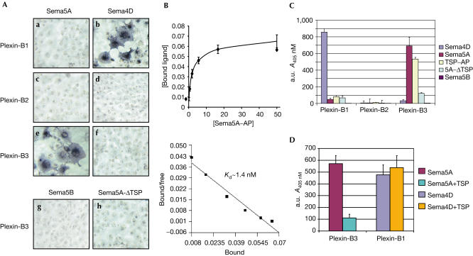
Semaphorins are a large family of molecular cues implicated in neural development and in a variety of functions outside the nervous system. Semaphorin 5A (Sema5A) is a transmembrane semaphorin, containing seven thrombospondin type-1 repeats, which was recently found to control axon guidance. Here we show that plexin-B3 is a high-affinity receptor specific for Sema5A. We further demonstrate that plexin-B3 activation by Sema5A mediates functional responses in plexin-B3-expressing cells (either fibroblasts, epithelial and primary endothelial cells). In addition, Sema5A can trigger the intracellular signalling of the hepatocyte growth factor/scatter factor receptor, Met, associated in a complex with plexin-B3. We thus conclude that Sema5A is able to elicit multiple functional responses through its receptor plexin-B3.


Figures

Figure 1
Figure 1
Sema5A binds to plexin-B3 with high affinity and specificity. (A) Binding of the indicated recombinant semaphorins fused to AP (Sema–AP) to COS cells expressing either plexin-B1 (a,b), plexin-B2 (c,d) or plexin-B3 (e–h). Cell-bound AP activity was revealed in situ using NBT–BCIP substrates, as described in Methods. (B) Scatchard analysis of Sema5A binding to cells expressing plexin-B3. COS cells expressing plexin-B3 or vector alone were incubated with increasing concentrations of Sema5A–AP for 1 h. Cell-bound semaphorin was then measured as AP activity on the chromogenic substrate pNPP, as described in Methods. The results are the mean of triplicate data points, and are representative of three independent experiments. (C) The binding of the indicated proteins (fused to AP) to cells expressing plexin-B1, plexin-B2 and plexin-B3 was quantified using the chromogenic substrate pNPP. Results are representative of three experiments performed in duplicate with consistent results. (D) Plexin-B3-expressing cells were incubated with Sema5A–AP alone (10 nM) or in the presence of equimolar amounts of soluble TSP-1 domain, and the binding was quantified as in (C). TSP-1 repeats markedly reduced the binding of Sema5A to plexin-B3, whereas they did not interfere with the interaction between Sema4D and its receptor plexin-B1.
Figure 2
Figure 2
Sema5A triggers the collapsing response in fibroblasts expressing plexin-B3. (A) NIH-3T3 fibroblasts expressing plexin-B3 were incubated with 30 nM of oligomerized Sema5A–Fc or Sema7A–Fc (see Methods). Plexin-B1-expressing cells were treated in the same conditions with oligomerized Sema5A–Fc or with Sema4D. Cells were stained with crystal violet and examined using a Leica DMIL microscope. (B) The collapsing response shown in (A) was quantified by photographing at least four representative × 20 microscope fields from two independent experiments and counting the number of cells that underwent contraction (defined as rounded cells having a diameter equal to or less than 20 μm; see Barberis et al, 2004). The results are the means of cell counts per field.
Figure 3
Figure 3
Sema5A triggers Met signalling by interaction with plexin-B3. (A) Western blotting of immunopurified endogenous Met from parental MLP-29 cells and cells engineered to express plexin-B3. Sema5A triggers Met activation and autophosphorylation in a plexin-B3-dependent manner. This is also indicated by a slight band shift of phosphorylated proteins, as expected. (B) MLP-29 cell migration assay using Transwell® inserts (see Methods). The indicated cell types were allowed to migrate towards oligomerized 10 nM Sema5A or 1 nM HGF in the presence of 5% FBS, or towards 20% FBS alone. After 16 h, migrated cells were fixed and stained with crystal violet. Cell migration was quantified by reading dye absorbance at 595 nm, and values were normalized to migration in control (ctr) medium (=100). Results are representative of three experiments performed in duplicate with consistent results. (C) Immunoblot analysis to demonstrate the expression of plexin-B3 (VSV-tagged) and Met in cells used for the experiments shown in (A,B). Met expression is barely detectable in cells subjected to targeted siRNA technology. (D) The chemotactic migration of primary HUVECs, either mock or infected to express plexin-B3, in response to either 4 nM HGF or 10 nM Sema5A added to the lower chamber of Transwell® insert. Cell migration was quantified as in (B). The data are the means of two independent experiments.

References

    1. Adams RH, Betz H, Puschel AW (1996) A novel class of murine semaphorins with homology to thrombospondin is differentially expressed during early embryogenesis. Mech Dev 57: 33–45 - PubMed
    1. Artigiani S, Comoglio PM, Tamagnone L (1999) Plexins, semaphorins, and scatter factor receptors: a common root for cell guidance signals? IUBMB Life 48: 477–482 - PubMed
    1. Artigiani S et al. (2003) Functional regulation of semaphorin receptors by proprotein convertases. J Biol Chem 278: 10094–10101 - PubMed
    1. Bahri SM, Chia W, Yang X (2001) Characterization and mutant analysis of the Drosophila sema 5c gene. Dev Dyn 221: 322–330 - PubMed
    1. Barberis D et al. (2004) Plexin signaling hampers integrin-based adhesion, leading to Rho-kinase independent cell rounding, and inhibiting lamellipodia extension and cell motility. FASEB J 18: 592–594 - PubMed

Publication types

MeSH terms