. 2004 May-Jun;51(3):267-81.
doi: 10.1111/j.1550-7408.2004.tb00567.x.

Morphologic and genetic variability in the marine planktonic ciliate Laboea strobila Lohmann, 1908 (Ciliophora, Oligotrichia), with notes on its ontogenesis

Affiliations

Morphologic and genetic variability in the marine planktonic ciliate Laboea strobila Lohmann, 1908 (Ciliophora, Oligotrichia), with notes on its ontogenesis

Sabine Agatha et al. J Eukaryot Microbiol. 2004 May-Jun.

Abstract

Laboea strobila Lohmann, 1908 is a conspicuous oligotrich ciliate in the marine plankton. In order to compare different populations, the morphology of specimens from the Mediterranean Sea, North Sea, and Irish Sea was investigated using live observation, protargol impregnation, and scanning electron microscopy. Furthermore, the PCR-amplified products of the SSrRNA gene from a monoclonal culture of L. strobila from the Mediterranean Sea were sequenced and aligned with sequences of other oligotrichs, including a population of L. strobila from the Atlantic coast of the USA. Finally, the data from the ecological literature were summarized and the cultivation methods were described. The SSrRNA gene sequences of the two distantly located L. strobila populations from the North Atlantic are identical. Likewise, the morphometrics of most populations so far investigated after protargol impregnation (i.e. from the North Atlantic) do not show obvious differences. In all computed phylogenetic trees, L. strobila groups with Strombidium species, forming a monophyletic taxon corresponding to the subclass Oligotrichia. These results are corroborated by the ontogenetic comparison. Since no type species was fixed for Laboea Lohmann, 1908, L. strobila was designated in the present paper.


Figures
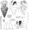
Fig. 1a–k
Fig. 1a–k
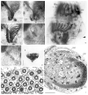
Laboea strobila, specimens from the Gulf of Trieste (a–e, g–k) and the Isle of Man (f) from life (a, d, h, k) and after protargol impregnation (b, c, e–g, i, j). a. A representative specimen showing the spiralled stripe of extrusomes accompanying the girdle kinety, except for the last half whorl. b, c. Ventral and dorsal views of same specimen. The girdle kinety performs ~4.5 turns around the cell, terminating dorsally near the posterior end (arrowhead). Although extrusomes and fibrillar structures are lacking in the last half kinety whorl, there is no change in kinetid structure and distance recognizable neither in protargol preparations nor in scanning electron micrographs (Fig. 3d, g–i). The macronuclear nodules are scattered throughout the cell. d. Pattern of cortical platelets. e, h. The resting extrusomes are deformed in protargol preparations (e), while acicular and ~ 24 × 0.7 μm in vivo (h). f. Pattern formed by extrusome attachment sites and the horizontal portion of an associated fibre. g. Dikinetidal girdle kinety with associated fibres. Arrowheads denote fibre portions extending underneath the following structure. i. Subjacent each anterior membranelle a row of argyrophilic granules extends. Their distal ends are connected by a C-shaped, ventrally open fibrillar structure. j. Anterior ventral cell portion. The paroral membrane probably comprises bare basal bodies as cilia are neither recognizable in protargol preparations nor in scanning electron micrographs (Fig. 2f, g). It is often accompanied by an additional longitudinal row of granules (arrowhead). k. Swimming trace. AM, anterior polykinetids; DC, distended cell surface; E, extrusomes/extrusome attachment sites; F, probably fibres; GK, girdle kinety; GR, granule rows; MA, macronuclear nodules; OF, oral fibres; PM, probably paroral membrane; VM, ventral polykinetids. Scale bars = 40 μm (a–c), 10 μm (h), 20 μm (j).
Fig. 2a–i
Fig. 2a–i
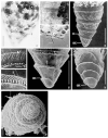
Laboea strobila, specimens from the Gulf of Trieste (a, c–i) and the Isle of Man (b) in the scanning electron microscope (a, b, d–h) and from life (c, i). a. Oblique ventral view. The ~1 μm long somatic cilia are arranged in a sinistrally spiralled girdle kinety underneath a stripe of extrusome attachment sites. b. Left lateral view showing the screw-like appearance of the cell due to the sinistrally spiralled girdle kinety. c. Live specimen. The dark inclusions are food vacuoles and sequestered plastids. d, f. Anterior ventral cell portions. Note the short cilia at the proximal and distal ends of each anterior membranelle (arrowheads). e. Top view showing the ventrally widely open zone of anterior membranelles and the separate ventral membranellar zone extending into the oral cavity. g. Oral cavity. Protargol preparations show the basal bodies of the paroral membrane. Instead of cilia, the SEM, however, reveals a ridge (arrowhead). h, i. Details of cell surface. Rhomboidal to polygonal cortical platelets cause the reticulation recognizable in scanning electron micrographs and compressed live cells. AM, anterior membranelles; AP, apical protrusion; BL, buccal lip; E, just exploding extrusomes; GK, girdle kinety; VM, ventral membranelles. Scale bars = 20 μm (a, b, d–f, i). 40 μm (c), 10 μm (g, h).
Fig. 3a–i
Fig. 3a–i
Laboea strobila, specimens from the Gulf of Trieste (a–g, i) and the Isle of Man (h) from life (a–c) and in the scanning electron microscope (d–i). a. Posterior cell portion of live specimen. The closely spaced extrusomes form a sinistrally spiralled stripe parallel to the girdle kinety. The cell is filled with fat droplets and sequestered plastids (asterisks). b, c. The resting extrusomes are acicular and up to 24 × 0.7 μm long. d, g–i. Lateral (d, g, h) and polar (i) views of posterior cell portion. The somatic cilia are arranged in a kinety, which commences underneath the buccal vertex and usually performs 4.5 sinistral spirals, terminating with an arc near the posterior end of dorsal side. The last kinety whorl is not accompanied by extrusomes and argyrophilic structures (probably fibres). A distinct gap, separating this kinety portion from the remaining ciliary row, is not recognizable. Its kinetids have the same distance like those of the remaining portion and protargol-impregnated cells do not reveal any change in the kinetid structure. Thus, a ventral kinety is not evident. e, f. Details of girdle kinety and stripe of extrusome attachment sites. The girdle kinety is composed of dikinetids each with a ~1 μm long cilium at the left basal body and a ciliary stub, forming a bulge, at the right (f; arrowheads); occasionally, the basal bodies and cilia are more closely spaced in the dorsal portion of the first kinety whorl, possibly in specimens commencing division (e). E, extrusomes/extrusome attachment sites; GK, girdle kinety. Scale bars = 20 μm (a–d, g–i), 2 μm (e, f.)
Fig. 4a–i
Fig. 4a–i
Laboea strobila, morphostatic specimens (a–c, h, i) and dividers (d–g) from the Gulf of Trieste after protargol impregnation (a–f) and from life (g), and cells from Sweden in the transmission electron microscope (h, i; unpubl. micrographs kindly provided by P. Jonsson). a–c. Buccal vertices with posterior portion of ventral membranellar zone. d, f. The oral primordium originates ventrally between the first and second kinety whorl left of midline. The formation of the new membranelles commences immediately.e. Dorsal view of a middle divider. Arrowhead denotes distended cell surface. g. Late divider. Arrowhead marks opisthe. h. Cross section of extrusomes. The extrusome structure is similar to that in other strombidiids, viz., a central lumen is surrounded by several concentric sheets and an electron-dense layer. Between the irregular compartment and the hexagonal electron-dense layer curved tubules extend (arrows). i. Oblique cross-section of anterior cell portion showing proximal ventral membranelles and arrangement of extrusomes. CP, cortical platelets; E, extrusomes; EL, electron-dense layer; F, probably fibres; FV, food vacuoles; GK, girdle kinety; GR, granule rows; L, extrusome lumen; OAM, opisthe’s anterior polykinetids; OF, oral fibres; OP, oral primordium; OPM, opisthe’s paroral membrane; OVM, opisthe’s ventral polykinetids; P, plastids; PAM, proter’s anterior polykinetids; SC, somatic cilium; VM, ventral polykinetids. Scale bars = 10 μm (a–d, f, i), 40 μm (e, g), 1 μm (h).
Fig. 5a–d
Fig. 5a–d
Laboea strobila, dividers from the Gulf of Trieste after protargol impregnation. a. Ventral view of an early divider. The oral primordium develops posterior to the left kinety portion, i.e. between the first and second kinety whorl on ventral side. The first membranelles immediately originate. b. Dorsal view of a middle divider. The formation of the membranelles has finished. The oral primordium shows an inverted orientation to the proter’s oral apparatus with the ventral membranelles and the paroral membrane (arrow) located dorsally. The minute breaks within the girdle kinety are likely preparation artifacts because not recognized in other middle dividers. Each macronuclear nodule shows a replication band. c. Ventral view of a late middle divider. The new oral apparatus evaginates between the first and second kinety whorl. The left girdle kinety portion is curved to surround the new membranellar zone posteriorly (arrow). Later, it separates from the right girdle kinety portion and forms the opisthe’s girdle kinety. In contrast to most hypotrichs and stichotrichs, the macronuclear nodules do not fuse before division but each elongates and divides alone, as indicated by the division furrows. d. Dorsal view of a late divider. The opisthe grows out, which apparently causes a shift of the new oral apparatus from the proter’s left to the right half of dorsal side. The girdle kinety of the proter commences mid-dorsally and extends sinistrally spiralled to the posterior cell end, while the opisthe’s girdle kinety already performs 1.5 turns. The structures (asterisk) at the centre of dorsal side are possibly newly forming extrusomes. Most macronuclear nodules have divided. DC, distended cell surface; GK, girdle kinety; OGK, opisthe’s girdle kinety; OP, oral primordium; PGK, proter’s girdle kinety; PM, paroral membrane; R, ridge. Scale bar = 40 μm.
Fig. 6
Fig. 6
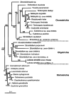
Phylogenetic tree for Laboea strobila computed with MrBayes ver. 2.01, employing the Bayesian Inference (Huelsenbeck and Ronquist, in press). The numbers are posterior probability values out of 32,300 trees.
Fig. 7a–i
Fig. 7a–i
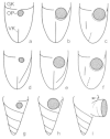
Schematized illustrations of the main ontogenetic events in three strombidiid genera. a–c. In Strombidium, the oral primordium develops underneath the girdle kinety (a). Later, the left portion of the girdle kinety curves along the posterior margin of the new, evaginating oral apparatus (b) and splits left laterally (c; based on Agatha 2003; Petz 1994; Song and Wang 1996). d-f. In Novistrombidium, the oral primordium originates above the left portion of the girdle kinety, which later surrounds the new, evaginating oral apparatus posteriorly and splits left laterally (e, f; based on Agatha 2003; Anigstein 1913). g-i. The ontogenesis of Laboea strobila is similar to that of Strombidium in the origin of the oral primordium underneath the left portion of the girdle kinety (g), which later curves posteriorly underneath the new, evaginating oral apparatus (h) and splits left laterally (i). The arrowhead marks the anterior end of the proter’s girdle kinety. Since Strombidium and Laboea recapitulate the ontogenetic behaviour of the girdle kinety in Novistrombidium, they are probably more derived. The oral primordium is represented by a shaded circle, the opisthe’s girdle kinety by a stippled line, and its ventral kinety by a dashed line. GK, girdle kinety; OP, oral primordium; VK, ventral kinety.

References

    1. Aescht E. Catalogue of the generic names of ciliates (Protozoa, Ciliophora) Denisia. 2001;1:1–350.
    1. Agatha S. Oligotrichea (aloricate species) In: Costello MJ, Emblow CS, White R, editors. European Register of Marine Species. A Check-list of the Marine Species in Europe and a Bibliography of Guides to Their Identification. 2001. pp. 42–44. (Patrimoines naturels). 50.
    1. Agatha S. Morphology and ontogenesis of Novistrombidium apsheronicum nov. comb. and Strombidium arenicola (Protozoa, Ciliophora): a comparative light microscopical and SEM study. Europ. J. Protistol. 2003;39:245–266.
    1. Alekperov I. Kh., Asadullayeva ES. New and little-known ciliates (orders Nassulida-Oligotrichida) from the Caspian Sea Apsheronian coast. Communication 2. Zool. Zh. 1997;76:1411–1417. (in Russian with English summary)
    1. Alekperov IH, Mamajeva NV. Planktonic infusoria from the Chuckchee and the Bering Seas. Zool. Zh. 1992;71:5–14. (in Russian with English summary)

Publication types

LinkOut - more resources