

Cell-surface peptidases
- PMID: 15219783
- PMCID: PMC7126636
- DOI: 10.1016/S0074-7696(04)35004-7
Cell-surface peptidases
Abstract
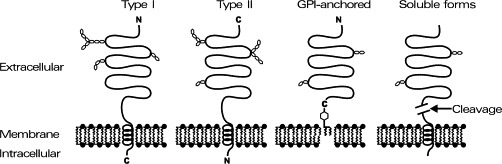
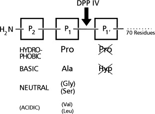
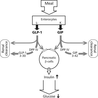
The cell surface has various functions: communicating with other cells, integrating into the tissue, and interacting with the extracellular matrix. Proteases play a key role in these processes. This review focuses on cell-surface peptidases (ectopeptidases, oligopeptidases) that are involved in the inactivation or activation of extracellular regulatory peptides, hormones, paracrine peptides, cytokines, and neuropeptides. The nomenclature of cell-surface peptidases is explained in relation to other proteases, and information is provided on membrane anchoring, catalytic sites, regulation, and, in particular, on their physiological and pharmacological importance. Furthermore, nonenzymatic (binding) functions and participation in intracellular signal transduction of cell surfaces peptidases are described. An overview on the different cell-surface peptidases is given, and their divergent functions are explained in detail. An example of actual pharmacological importance, dipeptidyl-peptidase IV (CD26), is discussed.
Figures
Similar articles
-
Human U937 cell surface peptidase activities: characterization and degradative effect on tumor necrosis factor-alpha.Eur J Immunol. 1992 Apr;22(4):923-30. doi: 10.1002/eji.1830220407. Eur J Immunol. 1992. PMID: 1348032
-
Membrane proteases in the bacterial protein secretion and quality control pathway.Microbiol Mol Biol Rev. 2012 Jun;76(2):311-30. doi: 10.1128/MMBR.05019-11. Microbiol Mol Biol Rev. 2012. PMID: 22688815 Free PMC article. Review.
-
Specificity of peptidases secreted by filamentous fungi.Bioengineered. 2018 Jan 1;9(1):30-37. doi: 10.1080/21655979.2017.1373531. Epub 2017 Sep 21. Bioengineered. 2018. PMID: 28857638 Free PMC article. Review.
-
Neuropeptidases and the metabolic inactivation of insect neuropeptides.Gen Comp Endocrinol. 2009 May 15;162(1):8-17. doi: 10.1016/j.ygcen.2008.12.011. Epub 2008 Dec 24. Gen Comp Endocrinol. 2009. PMID: 19135055 Review.
-
Post-proline-cleaving peptidases having DP IV like enzyme activity. Post-proline peptidases.Adv Exp Med Biol. 2000;477:103-9. doi: 10.1007/0-306-46826-3_10. Adv Exp Med Biol. 2000. PMID: 10849735 Review.
Cited by
-
B-Cell Lymphomas Secrete Novel Inhibitory Molecules That Disrupt HLA Class II-Mediated CD4+ T-Cell Recognition.Cells. 2025 Aug 7;14(15):1220. doi: 10.3390/cells14151220. Cells. 2025. PMID: 40801653 Free PMC article.
-
Decreased neprilysin and pulmonary vascular remodeling in chronic obstructive pulmonary disease.Am J Respir Crit Care Med. 2011 Feb 1;183(3):330-40. doi: 10.1164/rccm.201002-0154OC. Epub 2010 Sep 2. Am J Respir Crit Care Med. 2011. PMID: 20813891 Free PMC article.
-
Neuropeptide System Regulation of Prefrontal Cortex Circuitry: Implications for Neuropsychiatric Disorders.Front Neural Circuits. 2022 Jun 21;16:796443. doi: 10.3389/fncir.2022.796443. eCollection 2022. Front Neural Circuits. 2022. PMID: 35800635 Free PMC article. Review.
-
The protease complex consisting of dipeptidyl peptidase IV and seprase plays a role in the migration and invasion of human endothelial cells in collagenous matrices.Cancer Res. 2006 May 1;66(9):4652-61. doi: 10.1158/0008-5472.CAN-05-1245. Cancer Res. 2006. PMID: 16651416 Free PMC article.
-
Rainbow Trout (Oncorhynchus mykiss) as Source of Multifunctional Peptides with Antioxidant, ACE and DPP-IV Inhibitory Activities.Nutrients. 2023 Feb 6;15(4):829. doi: 10.3390/nu15040829. Nutrients. 2023. PMID: 36839187 Free PMC article.
References
-
- Abbott C.A., Baker E., Sutherland G.R., McCaughan G.W. Genomic organization, exact localization, and tissue expression of the human CD26 (dipeptidyl peptidase IV) gene. Immunogenetics. 1994;40:331–338. - PubMed
-
- Abbott C.A., Yu D.M., Woollatt E., Sutherland G.R., McCaughan G.W., Gorrell M.D. Cloning, expression and chromosomal localization of a novel human dipeptidyl peptidase (DPP) IV homolog, DPP8. Eur. J. Biochem. 2000;267:6140–6150. - PubMed
-
- Ahrén B., Simonsson E., Larsson H., Landin-Olsson M., Torgeirsson H., Jansson P.A., Sandqvist M., Bavenholm P., Efendic S., Eriksson J.W., Dickinson S., Holmes D. Inhibition of dipeptidyl peptidase IV improves metabolic control over a 4-week study period in type 2 diabetes. Diabetes Care. 2002;25:869–875. - PubMed
-
- Antczak C., De Meester I., Bauvois B. Transmembrane proteases as disease markers and targets for therapy. J. Biol. Regul. Homeost. Agents. 2001;15:130–139. - PubMed
Publication types
MeSH terms
Substances
LinkOut - more resources
Full Text Sources
Other Literature Sources
Miscellaneous
