. 2004 Sep 3;279(36):37789-97.
doi: 10.1074/jbc.M406225200. Epub 2004 Jun 27.

Mammalian wax biosynthesis. I. Identification of two fatty acyl-Coenzyme A reductases with different substrate specificities and tissue distributions

Affiliations

Mammalian wax biosynthesis. I. Identification of two fatty acyl-Coenzyme A reductases with different substrate specificities and tissue distributions

Jeffrey B Cheng et al. J Biol Chem. .

Abstract

The conversion of fatty acids to fatty alcohols is required for the synthesis of wax monoesters and ether lipids. The mammalian enzymes that synthesize fatty alcohols have not been identified. Here, an in silico approach was used to discern two putative reductase enzymes designated FAR1 and FAR2. Expression studies in intact cells showed that FAR1 and FAR2 cDNAs encoded isozymes that reduced fatty acids to fatty alcohols. Fatty acyl-CoA esters were the substrate of FAR1, and the enzyme required NADPH as a cofactor. FAR1 preferred saturated and unsaturated fatty acids of 16 or 18 carbons as substrates, whereas FAR2 preferred saturated fatty acids of 16 or 18 carbons. Confocal light microscopy indicated that FAR1 and FAR2 were localized in the peroxisome. The FAR1 mRNA was detected in many mouse tissues with the highest level found in the preputial gland, a modified sebaceous gland. The FAR2 mRNA was more restricted in distribution and most abundant in the eyelid, which contains wax-laden meibomian glands. Both FAR mRNAs were present in the brain, a tissue rich in ether lipids. The data suggest that fatty alcohol synthesis in mammals is accomplished by two fatty acyl-CoA reductase isozymes that are expressed at high levels in tissues known to synthesize wax monoesters and ether lipids.


Figures

SCHEME 1
SCHEME 1
Catalytic steps required to produce a wax monoester.
FIG. 1
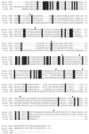
FIG. 1. Amino acid sequences of mouse, insect, and plant FAR enzymes
A, the deduced sequences of the proteins were aligned using the ClustalW (version 1.82) software program with identities indicated by black boxes. Amino acids are numbered on the right. Arrowheads above the mouse FAR1 sequence indicate the positions of 10 introns in the coding portion of the gene. The introns of the FAR2 gene were in the same positions. The GenBank™/EBI Data Bank accession numbers for the mouse FAR1 and FAR2 cDNA sequences are BC007178 and BC055759, respectively. Those for the human FAR1 and FAR2 cDNAs are AY600449 and BC022267, respectively.
FIG. 2
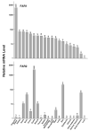
FIG. 2. Tissue distributions of mouse FAR1 and FAR2 mRNAs
The relative levels of each reductase mRNA were determined by real time PCR in the tissues and cell types indicated on the bottom of the figure using cyclophilin mRNA levels as a reference standard. The data for a given FAR mRNA were normalized to the threshold values (CT) determined in the liver (FAR1 mRNA = 27.4, FAR2 mRNA = 28.0) and then expressed on a log10 scale. This experiment was repeated twice on different days using the same preparations of tissue RNAs, which were isolated from pools of animals (organ samples) or dishes (cell samples).
FIG. 3
FIG. 3. Expression of mouse and human FAR enzymes in HEK 293 cells
Plasmid DNAs encoding the indicated FAR enzymes or a vector alone control (pCMV) were introduced into HEK 293 cells grown in 60-mm dishes. Approximately 24 h after transfection, fresh Dulbecco’s modified Eagle’s medium containing 33.3 μM BSA-conjugated palmitic acid and 2.4 μM BSA-conjugated [1-14C]palmitic acid was added to the cells. After an additional 24-h incubation, cells were washed once with PBS, harvested, and lipids extracted with chloroform:methanol in preparation for TLC. Chromatography on silica gel plates was performed for 0.5 h in solvent system 2, and the silica gel plate was exposed to Kodak BioMax MR film. The positions to which palmitate and hexadecanol standards migrated on the TLC plate are indicated on the right.
FIG. 4
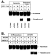
FIG. 4. Substrate and cofactor preferences of mouse FAR1 enzyme
A, insect cells (Sf9) in suspension were infected with baculovirus vectors encoding either a control protein (steroid 5β-reductase, 5β-Red) or the mouse FAR1 enzyme (mFAR1) for 48 h at a multiplicity of infection of 2–4. Cell membranes were prepared as described under “Experimental Procedures,” and aliquots (75 μg of protein) were incubated for 30 min at 37 °C in a reaction containing 2.5 mM β-NADPH and either 2.9 μM BSA-conjugated [1-14C]palmitic acid and 40 μM BSA-conjugated palmitic acid, with or without 1 mM ATP and 100 μM CoA, as indicated (lanes 1– 4), or 98 μM palmitoyl-CoA and 7 μM [1-14C]palmitoyl-CoA (lanes 5 and 6). The reaction was stopped by the addition of 100 μl of 6 N HCl, lipids were extracted and separated by TLC using solvent system 1, and radioactivity was detected by exposing the plate to x-ray film. The positions to which palmitate (substrate) and hexadecanol (product) migrated are shown on the right. B, Sf9 insect cells were infected with the indicated baculovirus vectors, and cell membranes were prepared. Aliquots (75 μg of protein) were incubated in reactions containing 98 μM palmitoyl-CoA, 7 μM [1-14C]palmitoyl-CoA, and 2.5 mM β-NADPH, 2.5 mM β-NADH, or 2.5 mM β-NADPH and 2.5 mM β-NADH, for 30 min at 37 °C. Lipids were extracted and analyzed as described in A. The experiments of A and B were repeated at least twice on separate days.
FIG. 5
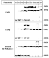
FIG. 5. Substrate preferences of mouse FAR1 and FAR2 enzymes
Baculovirus vectors encoding the fatty acyl-CoA reductase proteins or the control protein steroid 5β-reductase were used to infect adherent Sf9 cells at a multiplicity of infection of 5–10. Approximately 26 h after infection, intact cells were incubated with the indicated 14C-radiolabeled fatty acids conjugated to BSA. The final concentrations of fatty acids in the medium were 37.5 μM unlabeled lipid and 2.7–3 μM 1-14C-radiolabeled lipid. The cells were returned to the incubator for an additional 28 h, and thereafter lipids were extracted for TLC. The positions to which fatty alcohol (ROH), diacylglycerol (DAG), and monoacylglycerol (MAG) standards migrated on the plates developed in solvent system 1 are indicated on the right of the autoradiograms. Several radiolabeled compounds of unknown structure were present in the preparation of radiolabeled homo-γ-linolenic acid (20:3) used in these experiments, which were repeated two times.
FIG. 6
FIG. 6. Subcellular localization of mouse FAR1 and FAR2 enzymes
Chinese hamster ovary-K1 cells were transfected with the indicated expression vectors encoding nothing (pCMV), FLAG epitopetagged FAR1, or FLAG epitope-tagged FAR2 enzymes. After transient expression, cells were fixed, permeabilized, and incubated with a rabbit polyclonal antiserum directed against the SKL epitope of peroxisomal proteins (Anti-SKL) and a mouse monoclonal antibody directed against the FLAG epitope (Anti-FLAG). A, D, and G, detection of rabbit polyclonal antibodies directed against SKL epitope with rhodamine-conjugated goat anti-rabbit antiserum. B, E, and H, detection of mouse monoclonal antibody directed against FLAG epitope with fluorescein-conjugated goat anti-mouse antiserum. C, F, and I, merged images of rhodamine and fluorescein signals. Double indirect immunofluorescence microscopy was performed on a Zeiss 510 Laser Scanning Confocal microscope using a 63 × 1.3 NA PlanApo objective. The FLAG-FAR1 enzyme is detected in the peroxisome (middle panels). The FLAG-FAR2 enzyme is present in peroxisomes of cells expressing low levels of the protein and in the peroxisomes and endoplasmic reticulum of cells expressing high levels of the protein (bottom panels). These data are representative of two separate experiments.

References

    1. Downing DT, Strauss JS. J. Investig. Dermatol. 1974;62:228–244. - PubMed
    1. Nikkari T. J. Investig. Dermatol. 1974;62:257–267. - PubMed
    1. Jenks MA, Eigenbrode SD, Lemieux B. In: The Arabidopsis Book. Somerville CR, Meyerowitz EM, editors. American Society of Plant Biologists; Rockville, MD: 2002. doi/10.1199/tab.0016. http://www.aspb.org/publications/arabidopsis/
    1. Kunst L, Samuels AL. Prog. Lipid Res. 2003;42:51–80. - PubMed
    1. Wykle RL, Malone B, Snyder F. J. Lipid Res. 1979;20:890–896. - PubMed

Publication types