

The value of an elastic tissue stain in detecting venous invasion in colorectal cancer
- PMID: 15220375
- PMCID: PMC1770364
- DOI: 10.1136/jcp.2003.015826
The value of an elastic tissue stain in detecting venous invasion in colorectal cancer
Abstract
Background: Venous invasion by tumour is an independent prognostic indicator of both prognosis and risk of development of distant metastases in colorectal carcinoma. The use of special stains to aid its detection in pathology specimens is not currently universally recommended.
Aims: To determine whether an elastica stain significantly increases the incidence of detection of vascular invasion compared with routinely stained sections.
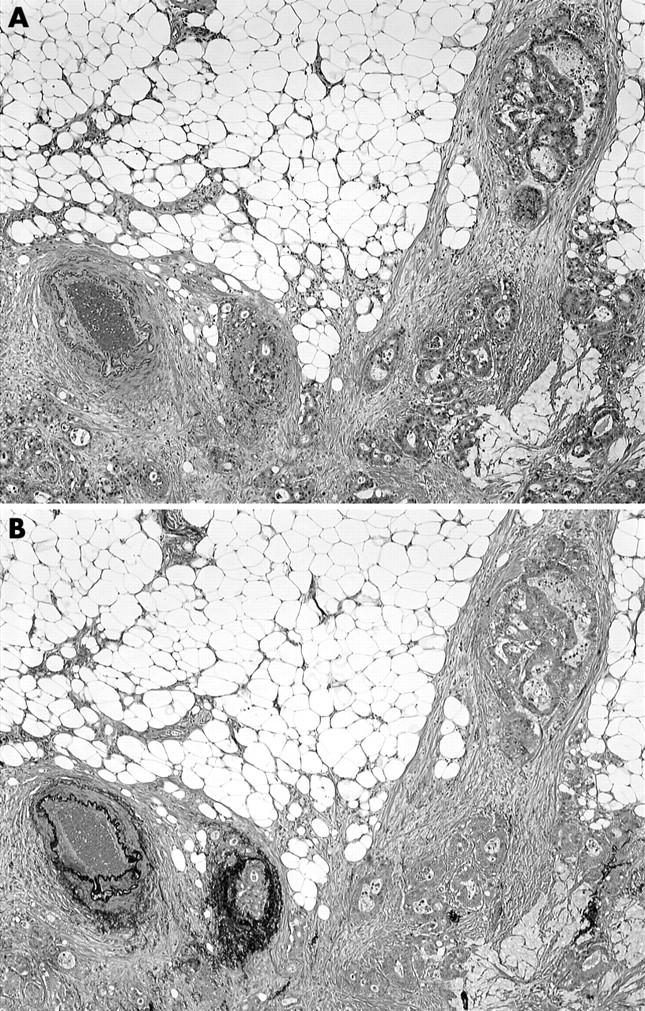
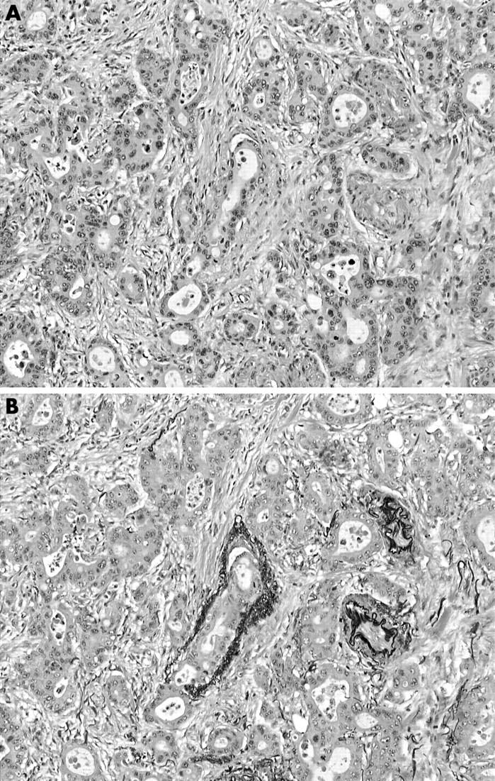
Methods: Serial sections from the 75 cases of colorectal carcinoma were stained by haematoxylin and eosin (H&E) only and elastica counterstained with H&E. The incidence of both intramural and extramural venous invasion was recorded and compared with that seen when the tumours were originally reported.
Results: Extramural venous invasion had been noted in 14 of the pathology reports and was seen in 18 cases when only the H&E sections were viewed in the study. It was present in 32 cases when elastica stained sections were analysed. Intramural venous invasion was seen in eight cases on H&E sections and 30 cases on elastica stained sections.
Conclusion: The use of elastica stained serial sections to detect venous invasion in tumours should be recommended in guidelines for the reporting of colorectal carcinomas.
Figures
Comment in
-
The benefits of elastic[corrected]--no stretch of the imagination [corrected].J Clin Pathol. 2006 Aug;59(8):888-9. doi: 10.1136/jcp.2005.032466. J Clin Pathol. 2006. PMID: 16873571 Free PMC article. No abstract available.
-
The prognostic benefits of routine staining with elastica to increase detection of venous invasion in colorectal cancer specimens.J Clin Pathol. 2011 Dec;64(12):1142. doi: 10.1136/jclinpath-2011-200284. Epub 2011 Aug 6. J Clin Pathol. 2011. PMID: 21821861 No abstract available.
References
-
- Brown CF, Warren S. Visceral metastasis from rectal carcinoma. Surg Gynaecol Obstet 1938;66:611–21.
-
- Talbot IC, Ritchie S, Leighton MH, et al. The clinical significance of invasion of veins by rectal cancer. Br J Surg 1980;67:439–42. - PubMed
-
- Krasna MJ, Flanbaum L, Cody RP, et al. Vascular and neural invasion in colorectal carcinoma. Cancer 1988;61:1018–23. - PubMed
-
- Shirouzu K , Isomoto H, Kakegawa T, et al. A prospective clinicopathological study of venous invasion in colorectal cancer. Am J Surg 1991;162:216–22. - PubMed
-
- Horn A , Dahl O, Morild I. Venous and neural invasion as predictors of recurrence in rectal adenocarcinoma. Dis Colon Rectum 1991;34:798–804. - PubMed
Publication types
MeSH terms
LinkOut - more resources
Full Text Sources
Medical