. 2004 Jul;78(14):7379-91.
doi: 10.1128/JVI.78.14.7379-7391.2004.

The N-terminal 85 amino acids of the barley stripe mosaic virus gammab pathogenesis protein contain three zinc-binding motifs

Affiliations

The N-terminal 85 amino acids of the barley stripe mosaic virus gammab pathogenesis protein contain three zinc-binding motifs

Jennifer N Bragg et al. J Virol. 2004 Jul.

Abstract

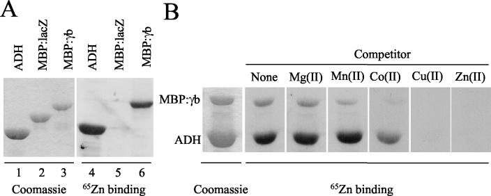
Barley stripe mosaic virus RNAgamma encodes gammab, a cysteine-rich protein that affects pathogenesis. Nine of the eleven cysteines are concentrated in two clusters, designated C1 (residues 1 to 23) and C2 (residues 60 to 85), that are arranged in zinc finger-like motifs. A basic motif (BM) rich in lysine and arginine (residues 19 to 47) resides between the C1 and C2 clusters. We have demonstrated that gammab binds zinc and that the C1, BM, and C2 motifs have independent zinc-binding activities. To evaluate the requirements for binding, mutations were introduced into each region. Cysteine residues at positions 7, 9, 10, 19, and 23 in the C1 motif were replaced with serines. In the BM, asparagines were substituted for lysines at positions 26 and 35, glutamine for arginine at position 25, and glycines for arginines at positions 33 and 36. The C2 mutations included cysteine replacements with serines at positions 60, 64, 71, and 81, and a histidine-to-leucine change at position 85. These mutations destroyed zinc-binding activity in each of the isolated motifs. gammab derivatives containing mutations in only two of the motifs retained the ability to bind zinc, whereas a gammab derivative containing mutations inactivating all three motifs destroyed the ability to bind zinc. Plants inoculated with transcripts containing combinations of the C1, BM, and C2 mutations elicited a "null" phenotype in barley characteristic of gammab deletion mutants and also delayed the appearance and reduced the size of local lesions in Chenopodium amaranticolor. These results show that zinc binding of each of the motifs is critical for the biological activity of gammab.


Figures

FIG. 1.
FIG. 1.
The tripartite genome of BSMV and subgenomic RNAs (sgRNAs) used for expression of the seven proteins encoded by the RNAs and motifs located within γb. (A) The filled circles represent the 5′ cap structure present on all of the BSMV genomic and subgenomic RNAs. Each 3′-proximal ORF terminates with a UAA codon, followed by an internal 4- to 40-nt polyadenylate sequence (An) that precedes the 238-nt tRNA-like 3′ terminus (solid rectangles). The three gRNAs, whose ORFs are illustrated by rectangular blocks, are designated α, β, and γ. RNAα encodes the αa protein, which is required for replication. The αa protein contains helicase (Hel) and methyltransferase (Mt) domains and forms the “helicase subunit” of the viral replicase complex. RNAβ encodes five proteins. The coat protein, βa, is translated directly from the gRNA. The overlapping triple gene block (TGB) proteins TGB1, TGB2, and TGB3 are each required for virus movement and are expressed from two subgenomic RNAs: sgRNAβ1 and sgRNAβ2. TGB1 contains a helicase (Hel) domain, whereas TGB2 and TGB3 are small hydrophobic proteins. TGB2′ is a minor translational readthrough protein that is dispensable for infection. RNAγ is bicistronic. The γa protein, which is translated from the gRNAγ, contains the GDD domain and is the polymerase subunit of the replicase. The cysteine-rich γb protein, which is expressed from the capped sgRNAγ, is involved in pathogenesis. (B) The γb protein is 152 amino acids in length and contains two cysteine-rich regions, C1 and C2, between amino acids 7 to 23 and 60 to 85, respectively. A BM, rich in lysine and arginine residues, lies between amino acids 19 to 47. Six heptad repeats that form a coiled-coil structure are predicted between amino acids 95 and 140.
FIG. 2.
FIG. 2.
Purification of γb by zinc affinity chromatography. Barley was inoculated with a derivative of BSMV in which the coat protein was not expressed, and infected tissue was harvested and extracted at 7 dpi. Protein samples were separated on 10% polyacrylamide gels, blotted onto nitrocellulose, and detected with γb and GAM-HRP antibodies. (A) The initial extract (I) that contains γb (lane 1) was subjected to zinc affinity chromatography. Lane 2 shows the γb protein that bound to the zinc affinity matrix (M), and lane 3 shows the effluent (E) fraction containing γb protein that failed to bind to the matrix. After application of the barley protein extract, the column was washed (W) with a low-salt buffer (lane 4). Lanes 5 to 7 show the γb protein present in the matrix, effluent, and wash fractions after the initial extract was applied to Sepharose matrix that was not charged with zinc. (B) Lane 1 shows the γb protein in the initial extract (I) from infected barley. Lanes 2 to 5 show the γb protein present in the effluent (E1), low-salt wash (W1), high-salt wash (H1), and matrix bound (M1) fractions after the first application to the zinc affinity matrix. Lanes 6 to 8 show the effluent (E2), low-salt wash (W2), and matrix-bound (M2) fractions of γb protein that were recovered from the E1 fraction after application to a fresh batch of zinc affinity matrix. (C) The γb protein bound to the zinc affinity matrix was eluted with a pH step gradient from pH 7.5 to 3.0.
FIG. 3.
FIG. 3.
Expression and subcellular distribution of wt γb and the γb(−)C1C2 mutant in barley. (A and B) Barley was inoculated with RNAα, the RNAβ B7 derivative in which the coat protein was not expressed, and an RNAγ encoding either the wt γb protein or the γb(−)C1C2 mutant and infected tissue was harvested at 7 dpi. (A) wt γb was extracted from infected tissue and fractionated. Lane 1, debris containing cellular and other fibrous materials that was retained after squeezing through cheese cloth; lane 2, the 1,000 × g P1 pellet fraction from expressed sap; lane 3, the 30,000 × g S30 soluble fraction; lane 4, the 30,000 × g P30 pellet fraction. (B) Lane 1, total wt γb protein recovered after grinding infected tissue directly in Laemmli buffer; lane 2, total γb(−)C1C2 protein extracted after grinding in Laemmli buffer; lane 3, γb(−)C1C2 cell debris retained by cheese cloth; lane 4, the γb(−)C1C2 1000 × g P1 pellet fraction from the cheesecloth filtrate; lane 5, the γb(−)C1C2 30,000 × g, S30 soluble fraction; lane 6, the γb(−)C1C2 30,000 × g, P30 pellet fraction. All fractions were separated on 10% SDS-polyacrylamide gels and blotted onto nitrocellulose. Proteins were detected with γb and GAM-HRP antibodies. Species of γb corresponding to sizes expected for monomers (M), dimers (D), tetramers (T), and aggregates (A) are designated by arrows. (C) Lane 1 (wt γb) and lane 2 [γb(−)C1C2] show [35S]methionine-labeled proteins recovered after translation in vitro in a rabbit reticulocyte system. Proteins were separated on a SDS-10% polyacrylamide gel, and the gel was dried and exposed to X-ray film.
FIG. 4.
FIG. 4.
65Zn(II)-binding activity of ADH and MBP-γb fusion proteins. Purified ADH and MBP fusion proteins separated on SDS-8% polyacrylamide gels were either stained with Coomassie brilliant blue and dried or blotted onto nitrocellulose, incubated with radiolabeled 65Zn(II), and exposed to X-ray film to assess zinc binding. (A) The ADH positive control, MBP-lacZ negative control, and MBP-γb proteins were tested for 65Zn(II) binding. The numbers at the bottom of the gel are for reference in the text. (B) Replicate samples of ADH and MBP-γb proteins were incubated either with 65ZnCl2 alone or with 65ZnCl2 and a divalent metal ion competitor present in a 1,000× molar concentration over the radioisotope.
FIG. 5.
FIG. 5.
65Zn(II)-binding activity of ADH and MBP-γb fusion proteins. (A and B) MBP fusion proteins separated on SDS-8% polyacrylamide gels were either stained with Coomassie brilliant blue and dried or blotted onto nitrocellulose, incubated with radiolabeled 65Zn(II), and exposed to X-ray film to assess zinc binding. The MBP fusion partner derivatives are designated at the top of each lane, and the numbers at the bottom are for reference in the text. The asterisk indicates the location of an E. coli-derived protein that copurifies with the MBP fusions. This protein is not visible in the Coomassie blue-stained gels but displays a minor zinc-binding signal.
FIG. 6.
FIG. 6.
Immunoblots of MBP fusion proteins. Purified MBP fusion proteins were separated on 8% SDS-polyacrylamide gels and blotted onto nitrocellulose. Proteins were detected with either MBP primary and goat anti-rabbit horseradish peroxidase secondary antibodies (top panels) or γb primary and GAM-HRP secondary antibodies (lower panels). Lane numbers are for reference in the text.
FIG. 7.
FIG. 7.
Symptoms elicited by BSMV containing either wt γb or γb zinc-binding mutant derivatives. The local lesion host C. amaranticolor (A) and the systemic host barley (B) were inoculated with transcripts of RNAα, RNAβ, and a RNAγ derivative encoding either the wt or a mutant γb protein. Leaves of healthy and infected plants were photographed between 6 and 11 dpi as designated. Before they were photographed at 11 dpi, the C. amaranticolor leaves that were inoculated with the wt virus abscised and became desiccated and shrunken. (C) RNA and proteins were extracted from barley tissue infected with BSMV expressing either wt γb or γb zinc-binding mutant derivatives. The γb derivatives are designated at the top of each lane. RNA was separated on a 1% agarose gel, blotted onto Nytran, and detected with a radiolabeled probe complementary to the 3′ region conserved among the BSMV RNAs. Proteins were separated on SDS-10% polyacrylamide gels, blotted onto nitrocellulose, and detected with the γb, coat protein, or TGB1 primary antibodies, respectively, and a GAM-HRP secondary antibody.

Similar articles

Cited by

References

    1. Auld, D. S. 2001. Zinc coordination sphere in biochemical zinc sites. Biometals 14:271-313. - PubMed
    1. Berg, J. M., and H. A. Godwin. 1997. Lessons from zinc-binding peptides. Annu. Rev. Biophys. Biomol. Struct. 26:357-371. - PubMed
    1. Chen, H., W. Kong, and R. Roos. 1995. The leader peptide of Theiler's murine encephalomyelitis virus is a zinc-binding protein. J. Virol. 69:8076-8078. - PMC - PubMed
    1. Donald, R. G. K., and A. O. Jackson. 1994. The barley stripe mosaic virus γb gene encodes a multifunctional cysteine-rich protein that affects pathogenesis. Plant Cell 6:1593-1606. - PMC - PubMed
    1. Donald, R. G. K., and A. O. Jackson. 1996. RNA-binding activities of barley stripe mosaic virus γb fusion proteins. J. Gen. Virol. 77:879-888. - PubMed

Publication types

Substances

LinkOut - more resources