. 2004 Jul;78(14):7455-64.
doi: 10.1128/JVI.78.14.7455-7464.2004.

Three novel cis-acting elements required for efficient plus-strand DNA synthesis of the hepatitis B virus genome

Affiliations

Three novel cis-acting elements required for efficient plus-strand DNA synthesis of the hepatitis B virus genome

Jehan Lee et al. J Virol. 2004 Jul.

Abstract

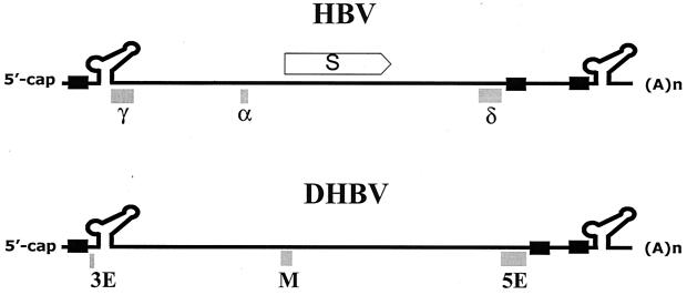
Synthesis of the relaxed-circular (RC) DNA genomes of hepadnaviruses by reverse transcriptase involves two template switches during plus-strand DNA synthesis. These template switches require repeat sequences (so-called donor and acceptor sites) between which a complementary strand of nucleic acid is transferred. To determine cis-acting elements apart from the donor and acceptor sites that are required for plus-strand RC DNA synthesis by hepatitis B virus (HBV), a series of mutants bearing a small deletion were made and analyzed for their impact on the viral genome synthesis. We found three novel cis-acting elements in the HBV genome: one element, located in the middle of the minus strand, is indispensable, whereas the other two elements, located near either end of the minus strand, contribute modestly to the plus-strand RC DNA synthesis. The data indicated that the first element facilitates plus-strand RNA primer translocation or subsequent elongation during plus-strand RC DNA synthesis, while the last two elements, although distantly located on the minus strand, act at multiple steps to promote plus-strand RC DNA synthesis. The necessity of multiple cis-acting elements on the minus-strand template reflects the complex nature of hepadnavirus reverse transcription.


Figures

FIG. 1.
FIG. 1.
Model for hepadnavirus reverse transcription. (A) Initiation of minus-strand DNA synthesis. The thin line represents the pgRNA. The direct repeats, DR1 and DR2, are indicated by boxes. Minus-strand DNA synthesis is templated by the UUCA sequence within the bulge region of the 5′ ɛ. The oval circle represents the P protein. (B) Minus-strand template switch. The 4-nt-linked viral P protein translocates to an acceptor site, the UUCA sequence, overlapping the 3′ copy of DR1 via 4-bp homology. (C) Elongation and completion of minus-strand DNA synthesis. Following the minus-strand transfer, minus-strand synthesis resumes, with concomitant degradation of the pgRNA by RNase H activity encoded by the P protein. (D) In situ priming from DR1. Some of the plus-strand primers do not translocate but are used to initiate plus-strand synthesis from DR1 to generate a DL DNA. (E) Generation of a DL genome. (F) Plus-strand primer translocation to DR2. The RNA primer contains a DR1 sequence complementary to DR2. This complementarity is required for subsequent translocation to DR2. To generate the circular duplex genome, the RNA primer translocates from DR1 to an acceptor site, DR2. (G) Initiation of plus-strand DNA synthesis following translocation. After translocation to DR2, plus-strand DNA synthesis is initiated at DR2. (H) Template switch to circularize the viral genome. The growing point of plus-strand DNA synthesis switches templates from the 5′ end to the 3′ end of minus-strand DNA. (I) Generation of an RC DNA genome.
FIG. 2.
FIG. 2.
Maps of deletion mutants used in this study. (A) Map of the pgRNA of HBV. The structure of pgRNA is shown with cis-acting elements that are known to be essential for viral genome synthesis; R represents the region of terminal redundancy that includes DR1 and ɛ. The direct repeats, DR1 and DR2, are represented as boxes; the 3′ copy of DR1 is designated DR1*. The encapsidation signal, ɛ, is indicated by the stem-loop structure at the 5′ end of the pgRNA. ORFs for C (core) and P (polymerase) are indicated by boxes. (B) A series of deletion mutants that encompass the genome from 5′ ɛ to DR2 were made. The size of the deletion in each mutant (in nucleotides) is indicated in parentheses after the plasmid name. The nucleotide positions of each deletion mutant are indicated by a line along with nucleotide number, inclusive. Most of the deletion mutants used in this study are defective in the expression of functional C or P protein, which are instead provided in trans by a helper plasmid. None of these deletion mutants exhibited a dominant negative effect on viral genome replication (data not shown).
FIG. 3.
FIG. 3.
Southern analysis reveals three novel cis-acting elements in the viral genome. (A) Southern analysis of the replication intermediate DNAs isolated from cytoplasmic core particles from cells transfected by each deletion mutant along with a helper plasmid providing C and P proteins. The positions of RC, DL, and single-stranded (SS) DNA forms are indicated. The viral DNAs extracted from HepG2.2.15 cells (an HBV-producing cell line) served as size markers for the three replication intermediates (23). WT, wild type. (B) Relative amount of RC DNA with respect to RC plus DL DNA. The amounts of RC DNA and DL DNA for each mutant as determined from at least six independent transfections were quantified with a Bio-imaging analyzer. The mean ratio and standard deviation for each mutant are shown.
FIG. 4.
FIG. 4.
Strategies used to measure replication intermediates by primer extension analysis. Only part of the plus-strand DNA of the RC genome is shown on the top for clarity; DR1, DR2, and r are boxed or underlined. The positions of the annealing sites for the primers are indicated by arrows, with the nucleotide numbers in parentheses. Four replication intermediates found in cytoplasmic capsids are shown. Each replication intermediate has a full-length minus-strand DNA (thick line) with the P protein (oval) linked to the 5′ end. (A) Single-stranded DNA. The M primer is designed to map the 5′ end of the minus-strand DNA. (B) DL DNA. The DL DNA is generated when the RNA primer initiates synthesis at its site of generation (so-called in situ priming). The P primer is designed to map the 5′ end of the plus-strand DNA. (C) RC DNA before circularization. The RNA primer for the plus-strand synthesis anneals to the DR2 site on the minus strand. Primer extension with the B primer will yield a signal at DR2, while that with the P primer will not. (D) RC DNA after circularization. The B primer is designed to map the 5′ end of the plus-strand DNA before circularization, whereas the P primer is designed to map the 5′ end of the plus-strand DNA after circularization.
FIG. 5.
FIG. 5.
Primer extension analysis reveals that three regions (the α, γ, and δ elements) in the minus-strand DNA facilitate primer translocation to DR2 during plus-strand DNA synthesis. (A) Primer extension analysis to determine the levels and positions of the 5′ termini of plus-strand DNA for the deletion mutants indicated. The P primer was used to determine the levels and positions of the 5′ ends of the plus-strand DNAs located at DR2 and DR1 for the mutant and wild-type (WT) viruses. A sequencing ladder generated with the P primer flanks the primer extension reactions. Positions of DR1 and DR2 are indicated. (B) Detection of 5′ termini of minus-strand DNA. The M primer was used to determine the level and position of the 5′ end of minus-strand DNA. A sequencing ladder made with primer M is shown for comparison. (C) The amount of plus-strand DNA initiated at DR2 relative to the amount of minus-strand DNA initiated at DR1 was calculated for each mutant after normalization to the extension products detected from the internal standard by the M primer. For each mutant, the mean and standard deviation of the ratio of plus-strand DNA to minus-strand DNA from three independent transfections is shown. (D) The amount of plus-strand DNA initiated by in situ priming relative to the amount of minus-strand DNA was calculated for each mutant. Other details are the same as for panel C.
FIG. 6.
FIG. 6.
Detection of plus-strand DNA before circularization. (A) The B primer was used to determine the level and position of the 5′ end of plus-strand DNA before circularization. The position of DR2 is indicated. A sequencing ladder made with the B primer is shown for comparison. (B) The amount of plus-strand DNA initiated at DR2 relative to the amount of minus-strand DNA initiated at DR1 was calculated for each mutant after normalization as described for Fig. 5C. For each mutant, the mean and standard deviation of the ratio of plus-strand DNA to minus-strand DNA from three independent transfections is shown. WT, wild type.
FIG. 7.
FIG. 7.
Determination of the boundary of the α region by deletion analysis. (A) Map of the five small deletion mutants within the α region. The nucleotide sequences deleted in each mutant are shown by solid line along with the nucleotide number, inclusive. The size of the deletion in each mutant (in nucleotides) is indicated in parentheses after the plasmid name. (B) Southern blot analysis was performed as described for Fig. 3A. Each mutant generated in the background of a P-null version of the wild type was transfected into cells along with the helper. WT, wild type; SS, single-stranded DNA; S/M, size marker.
FIG. 8.
FIG. 8.
Comparison of cis-acting elements required for RC DNA synthesis by HBV with those required for RC DNA synthesis by DHBV. The direct repeats, DR1 and DR2, are not drawn to scale. The S (surface antigen) ORF is shown for comparison. The nucleotide sequence corresponding to 3E of DHBV, which is positioned between the 5′ copy of DR1 and 5′ ɛ, was not examined in this work.

Similar articles

Cited by

References

    1. Condreay, L. D., T. T. Wu, C. E. Aldrich, M. A. Delaney, J. Summers, C. Seeger, and W. S. Mason. 1992. Replication of DHBV genomes with mutations at the sites of initiation of minus- and plus-strand DNA synthesis. Virology 188:208-216. - PubMed
    1. Donello, J. E., A. A. Beeche, G. J. Smith III, G. R. Lucero, and T. J. Hope. 1996. The hepatitis B virus posttranscriptional regulatory element is composed of two subelements. J. Virol. 70:4345-4351. - PMC - PubMed
    1. Galibert, F., E. Mandart, F. Fitoussi, P. Tiollais, and P. Charnay. 1979. Nucleotide sequence of the hepatitis B virus genome (subtype ayw) cloned in E. coli. Nature 281:646-650. - PubMed
    1. Ganem, D., and R. Schneider. 2001. Hepadnaviridae: the viruses and their replication, 4th ed., vol. 2. Lippincott-Raven Publishers, Philadelphia, Pa.
    1. Goff, S. 2001. Retroviridae: the retroviruses and their replication, p. 1871-1939. In P. M. Hawley and D. M. Knipe (ed.), Field's virology, 4th ed. Lippincott-Raven Publishers, Philadelphia, Pa.

Publication types

LinkOut - more resources