. 2004 Jul;78(14):7508-17.
doi: 10.1128/JVI.78.14.7508-7517.2004.

Cellular and viral requirements for rapid endocytic entry of herpes simplex virus

Affiliations

Cellular and viral requirements for rapid endocytic entry of herpes simplex virus

Anthony V Nicola et al. J Virol. 2004 Jul.

Abstract

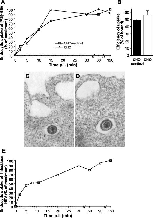
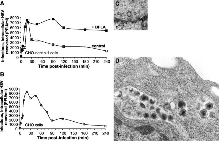
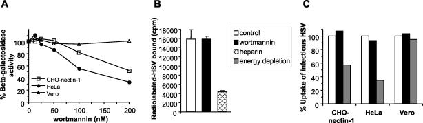
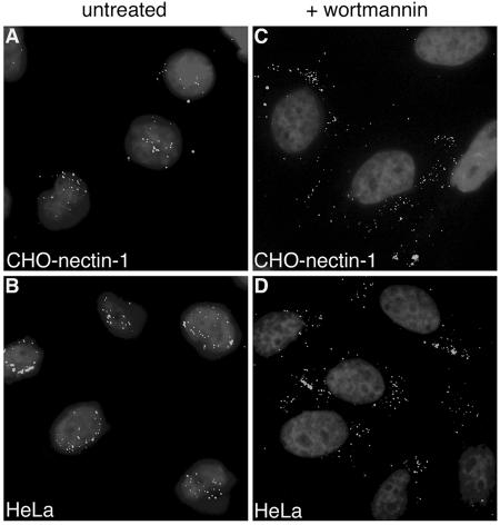
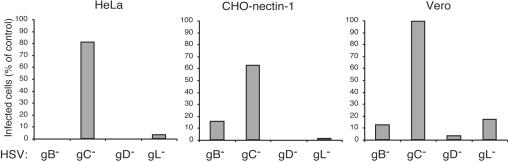
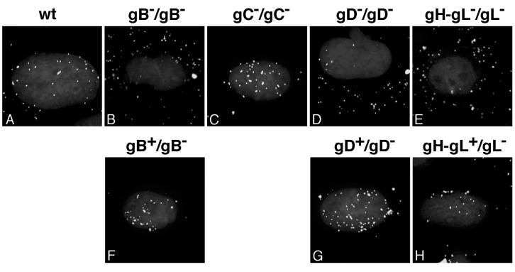
It was recently demonstrated that herpes simplex virus (HSV) successfully infects Chinese hamster ovary (CHO) cells expressing glycoprotein D (gD) receptors and HeLa cells by an endocytic mechanism (A. V. Nicola, A. M. McEvoy, and S. E. Straus, J. Virol. 77:5324-5332, 2003). Here we define cellular and viral requirements of this pathway. Uptake of intact, enveloped HSV from the cell surface into endocytic vesicles was rapid (t(1/2) of 8 to 9 min) and independent of the known cell surface gD receptors. Following uptake from the surface, recovery of intracellular, infectious virions increased steadily up to 20 min postinfection (p.i.), which corresponds to accumulation of enveloped virus in intracellular compartments. There was a sharp decline in recovery by 30 min p.i., suggesting loss of the virus envelope as a result of capsid penetration from endocytic organelles into the cytosol. In the absence of gD receptors, endocytosed virions did not successfully penetrate into the cytosol but were instead transported to lysosomes for degradation. Inhibitors of phosphatidylinositol (PI) 3-kinase, such as wortmannin, blocked transport of incoming HSV to the nuclear periphery and virus-induced gene expression but had no effect on virus binding or uptake. This suggests a role for PI 3-kinase activity in trafficking of HSV through the cytosol. Viruses that lack viral glycoproteins gB, gD, or gH-gL were defective in transport to the nucleus and had reduced infectivity. Thus, similar to entry via direct penetration at the cell surface, HSV entry into cells by wortmannin-sensitive endocytosis is efficient, involves rapid cellular uptake of viral particles, and requires gB, gD, and gH-gL.


Figures

FIG. 1.
FIG. 1.
Kinetics of HSV uptake by endocytosis and role of gD receptors. (A) Role of nectin-1 in uptake from the cell surface. 35S-labeled virus was added to CHO cells, which lack any known gD receptors, or CHO-nectin-1 cells for 2 h at 4°C. Following shift to 37°C for the time indicated as minutes p.i., nonendocytosed virus was removed by treatment with 2 mg of proteinase K/ml or cells were mock treated to determine the amount of total virus bound. The fraction of total cell-associated cpm that is protected from protease treatment reflects HSV that has been endocytosed. Uptake at time zero was set to 0%, and maximum uptake was set to 100%. Each value is the mean of triplicate determinations. (B) Efficiency of uptake. Uptake of radiolabeled HSV at 30 min p.i. was determined as described for panel A. Efficiency is defined here as protease-resistant cpm out of cpm bound. Shown are the mean values of triplicate samples with standard error. Data are representative of four similar experiments. EM analysis of HSV uptake by CHO-nectin-1 (C) cells or CHO cells (D). HSV-1 KOS (MOI of 50) was bound to cells for 2 h at 4°C followed by a shift to 37°C for 3 min, and then cells were processed for EM. Magnification, ×90,000. Enveloped virions are 150 to 200 nm in diameter. (E) Uptake of infectious HSV from the cell surface. HSV-1 KOS was bound to CHO-nectin-1 cells for 2 h at 4°C (MOI of 0.5). Cells were washed with PBS and incubated at 37°C, and extracellular virus was inactivated by acid treatment at the indicated times. At 8 h p.i. cells were fixed and random fields of 400 to 500 cells per sample were evaluated. Total cell number was evaluated by nuclear staining with DAPI, and infected cells were detected by immunofluorescence with an anti-HSV polyclonal antibody. Maximum infectivity was set to 100%.
FIG. 2.
FIG. 2.
Intracellular trafficking of HSV. HSV-1 KOS was bound to CHO-nectin-1 cells (A) or CHO cells (B) for 2 h at 4°C. Cultures were shifted to 37°C in the presence or absence of 200 nM BFLA. At the indicated time p.i., following inactivation of extracellular virus, cells were lysed by two cycles of freezing and thawing. The numbers of infectious, intracellular particles were assayed by plaque titration on Vero cells. Data shown are representative of at least three independent experiments. For ultrastructural analysis, HSV-1 KOS (MOI of 50) was bound to cells for 2 h at 4°C followed by a shift to 37°C for 1 h, and then samples were processed for EM. (C) Naked, empty viral capsid docked at the nuclear pore complex of a CHO-nectin-1 cell. (D) Accumulation of degraded HSV particles in CHO cells. Enveloped virions are 150 to 200 nm, and capsids are ∼100 nm in diameter. Magnification, 90,000×. n, nucleus; ne, nuclear envelope.
FIG. 3.
FIG. 3.
Effect of wortmannin on HSV entry. (A) CHO-nectin-1, HeLa, or Vero cells were pretreated with the indicated concentrations of wortmannin for 30 min. Cells were infected with the HSV-1 strain KOS (for CHO-nectin-1 cells) or lacZ+ KOS 7134 (for HeLa and Vero cells) at an MOI of 1 for 6 h in the continued presence of agent. Entry was measured as the percentage of β-galactosidase activity relative to that obtained in the absence of wortmannin. (B) Effect of wortmannin on virus binding to cells. 35S-labeled HSV was added to CHO-nectin-1 cells (MOI of 3) for 2 h at 4°C in the presence of 200 nM wortmannin or 100 μg of heparin/ml. Cells were washed three times, and then cell-associated cpm of detergent lysates were determined. Each value is the mean and standard deviation of triplicate determinations. The legend applies to both panels B and C. (C) Effect of wortmannin on uptake of infectious virus. HSV was bound to CHO-nectin-1, HeLa, or Vero cells at 4°C for 2 h. Cells were washed and then treated for 15 min at 4°C with medium containing 200 nM wortmannin or with sodium azide and 2-deoxy-d-glucose (energy depletion medium). Cultures were shifted to 37°C in the presence of these agents for 1 h to allow virus to be taken up by endocytosis (CHO-nectin-1 and HeLa cells) or to penetrate directly from the surface (Vero cells). Inhibitor was removed, and extracellular virus was acid inactivated. Cultures were returned to 37°C for 7 h in normal medium without inhibitors. Cells were fixed, and random fields of 400 to 500 cells were evaluated per sample. Total cell number was evaluated by nuclear staining with DAPI, and infected cells were detected by immunofluorescence with an anti-HSV polyclonal antibody. One-hundred percent uptake was taken as the level of infection detected in control untreated cultures. Each value represents the mean of two or three independent experiments.
FIG. 4.
FIG. 4.
Effect of PI 3-kinase inhibitors on the cellular distribution of incoming virions. HSV-1 KOS K26GFP (MOI of 20) was bound to CHO-nectin-1 (A and C) or HeLa (B and D) cells for 2 h at 4°C. Cells were washed with PBS and then warmed in medium containing no agent (A and B) or 10 nM wortmannin (C and D). Infection proceeded for 2.5 h in the presence of cycloheximide. Cells were washed with PBS and fixed in 3% paraformaldehyde. Nuclei were counterstained with DAPI. Punctate fluorescence indicates HSV particles. Cells were viewed with a 63× oil immersion objective.
FIG. 5.
FIG. 5.
HSV glycoprotein requirements for infection of cells that support distinct entry pathways. HeLa, CHO-nectin-1, or Vero cells were infected with HSV-1 strain KOS mutants devoid of gB, gC, gD, or gL for 10 h. Total cell number and HSV antigen-positive cells were quantitated by immunofluorescence microscopy. Four-hundred to 500 cells per sample were evaluated. One-hundred percent represents the infectivity of an equivalent number of particles of the corresponding complemented virus.
FIG. 6.
FIG. 6.
HSV glycoprotein requirements for transport of particles to the nuclear periphery. CHO-nectin-1 cells were infected with ∼108 particles of HSV-1 wild-type (wt) KOS (A), gB null (B), gC null (C), gD null (D), gH-gL null (E), complemented gB null (F), complemented gD null (G), or complemented gH-gL null (H) for 2.5 h in the presence of cycloheximide. The envelope content/genome content of the infecting virus is indicated above each panel. Following fixation, cells were probed with anti-HSV VP5 antibody and Alexa Fluor 488-conjugated secondary antibody to visualize individual particles. Nuclei were counterstained with DAPI. Cells were viewed with a 100× oil immersion objective.

Similar articles

Cited by

References

    1. Akula, S. M., P. P. Naranatt, N. S. Walia, F. Z. Wang, B. Fegley, and B. Chandran. 2003. Kaposi's sarcoma-associated herpesvirus (human herpesvirus 8) infection of human fibroblast cells occurs through endocytosis. J. Virol. 77:7978-7990. - PMC - PubMed
    1. Amici, C., G. Belardo, A. Rossi, and M. G. Santoro. 2001. Activation of I kappa B kinase by herpes simplex virus type 1. A novel target for anti-herpetic therapy. J. Biol. Chem. 276:28759-28766. - PubMed
    1. Batterson, W., D. Furlong, and B. Roizman. 1983. Molecular genetics of herpes simplex virus. VIII. Further characterization of a temperature-sensitive mutant defective in release of viral DNA and in other stages of the viral reproductive cycle. J. Virol. 45:397-407. - PMC - PubMed
    1. Cai, W., and P. A. Schaffer. 1991. A cellular function can enhance gene expression and plating efficiency of a mutant defective in the gene for ICP0, a transactivating protein of herpes simplex virus type 1. J. Virol. 65:4078-4090. - PMC - PubMed
    1. Cai, W. H., B. Gu, and S. Person. 1988. Role of glycoprotein B of herpes simplex virus type 1 in viral entry and cell fusion. J. Virol. 62:2596-2604. - PMC - PubMed