. 2004 Jul;78(14):7653-66.
doi: 10.1128/JVI.78.14.7653-7666.2004.

Replication of herpes simplex virus 1 depends on the gamma 134.5 functions that facilitate virus response to interferon and egress in the different stages of productive infection

Affiliations

Replication of herpes simplex virus 1 depends on the gamma 134.5 functions that facilitate virus response to interferon and egress in the different stages of productive infection

Xianghong Jing et al. J Virol. 2004 Jul.

Abstract

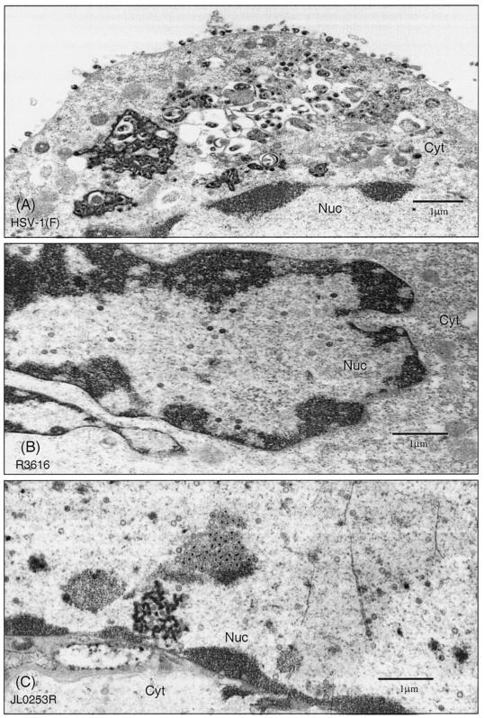
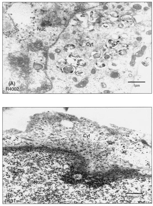
The ability of the gamma(1)34.5 protein to suppress the PKR response plays a crucial role in herpes simplex virus pathogenesis. In this process, the gamma(1)34.5 protein associates with protein phosphatase 1 to form a large complex that dephosphorylates eIF-2alpha and thereby prevents translation shutoff mediated by PKR. Accordingly, gamma(1)34.5 null mutants are virulent in PKR-knockout mice but not in wild-type mice. However, gamma(1)34.5 deletion mutants, with an extragenic compensatory mutation, inhibit PKR activity but remain avirulent, suggesting that the gamma(1)34.5 protein has additional functions. Here, we show that a substitution of the gamma(1)34.5 gene with the NS1 gene from influenza A virus renders viral resistance to interferon involving PKR. The virus replicates as efficiently as wild-type virus in SK-N-SH and CV-1 cells. However, in mouse 3T6 cells, the virus expressing the NS1 protein grows at an intermediate level between the wild-type virus and the gamma(1)34.5 deletion mutant. This decrease in growth, compared to that of the wild-type virus, is due not to an inhibition of viral protein synthesis but rather to a block in virus release or egress. Virus particles are predominantly present in the nucleus and cytoplasm. Notably, deletions in the amino terminus of the gamma(1)34.5 protein lead to a significant decrease in virus growth in mouse 3T6 cells, which is independent of eIF-2alpha dephosphorylation. In correlation, a series of deletions in the amino-terminal domain impair nuclear as well as cytoplasmic egress. These results indicate that efficient viral replication depends on the gamma(1)34.5 functions required to prevent the PKR response and to facilitate virus egress in the different stages during virus infection.


Figures

FIG. 1.
FIG. 1.
(A) Schematic representation of the genome structure of HSV-1 and its derivatives. The two covalently linked components of HSV-1 DNA, L and S, each consist of unique sequences, UL and US, respectively, flanked by inverted repeats (41). The reiterated sequences flanking UL, designated as ab and ba′, are each 9 kbp in size, whereas the repeats flanking US, designated ac′ and ca, are 6.3 kbp in size. The location of the γ134.5 gene is shown in the expanded portions of the inverted repeat sequences b and b′. Since the b sequence is repeated in an inverted orientation, there are two copies of the γ134.5 gene per genome. HSV-1(F) is the prototype strain used in our laboratory (24). In R3616, the coding region between BstEII-StuI sites of the γ134.5 gene is deleted (21). In recombinant virus JL0253R, the γ134.5 gene is replaced with the influenza A virus NS1 gene (43), which is driven by a cytomegalovirus promoter. Restriction site designations are as follows: N, NcoI; Be, BstEII; S, SacI; and St, StuI. (B) Autoradiographic images of viral DNAs. Vero cells were infected with the indicated viruses at 10 PFU per cell. At 18 h postinfection, cells were harvested, and viral DNA was prepared and then digested with either BamHI, EcoRI and XhoI, or BstEII and DraIII. Samples were electrophoretically separated on 0.8% agarose gels and transferred to a nitrocellulose membrane. The tk gene was detected by hybridization to a 32P-labeled BamHI Q fragment of HSV-1. Similarly, the NS1 gene was probed with a 32P-labeled NS1 fragment spanning nucleotides 45 to 652, and the γ134.5 gene was probed with a 32P-labeled γ134.5 fragment spanning nucleotides 345 to 579. (C) Expression of the γ134.5 protein and the NS1 protein. Vero cells were either mock infected or infected with the indicated viruses at 10 PFU per cell. At 18 h postinfection, the cells were harvested and subjected to electrophoresis, transferred to a nitrocellulose membrane, and reacted with either anti-NS1 antibody or anti-γ134.5 antibody.
FIG. 2.
FIG. 2.
Viral response to alpha interferon. Monolayers of Vero cells were either untreated or pretreated with human leukocyte alpha interferon (1,000 U/ml; Sigma) for 20 h. Cells were then infected with viruses at 0.05 PFU per cell and incubated at 37°C. At 48 h postinfection, cells were harvested, and virus yields were determined on Vero cells (14). Data represent the average from three independent experiments, with the standard deviation indicated.
FIG. 3.
FIG. 3.
Growth properties of wild-type HSV-1(F), the γ134.5 deletion mutant, and the recombinant virus expressing the NS1 protein. Confluent monolayers of SK-N-SH (A), CV-1 (B), and MEF 3T6 (C) cells were infected with HSV-1(F), R3616, or JL0253R at 0.01 PFU per cell and incubated at 37°C. Viruses were harvested at 24 and 48 h postinfection. Samples were freeze-thawed three times and titrated on Vero cells at 37°C. As a parallel experiment, confluent monolayers of MEF 3T6 cells were also infected at 10 PFU per cell (D). Viruses were harvested at 24 h postinfection and titrated as described above. The data represent an average from three independent experiments, and the error bars indicate standard deviations. MOI, multiplicity of infection.
FIG. 4.
FIG. 4.
(A) Synthesis of viral proteins in virus-infected MEF 3T6 cells. Confluent monolayers of MEF 3T6 cells were either mock infected or infected with the indicated viruses at 10 PFU per cell. At 16 h postinfection, cells were harvested, solubilized, subjected to polyacrylamide gel analysis, transferred to a nitrocellulose sheet, and reacted with polyclonal antibodies against whole HSV-1 antigens (Dako Corporation). (B) Phosphorylation state of eIF-2α. The same membrane described above (A) was stripped and probed with antibodies against eIF-2α and phosphorylated eIF-2α (Cell Signaling Technology). The positions of eIF-2α and phosphorylated eIF-2α are shown on the right.
FIG. 5.
FIG. 5.
Cellular distribution of virus particles in MEF 3T6 cells. Confluent monolayers of MEF 3T6 cells were infected with HSV-1(F), R3616, or JL0253R at 0.5 PFU per cell. At 24 h postinfection, cells were first fixed in 4% glutaraldehyde in 100 mM phosphate buffer (pH 6.8 to 7.2) and then fixed in 1% osmium tetroxide. Cells were dehydrated in ethanol, embedded in LX112 resin, and stained with uranyl acetate and lead citrate. Thin sections were prepared and viewed with a Joel 1220 transmission electron microscope at 80 kV. Images were captured with a Gatan digital CCD camera. (A) HSV-1(F)-infected MEF 3T6 cells. (B) R3616-infected MEF 3T6 cells. (C) JL0253R-infected MEF 3T6 cells. Scale bars are shown in each panel. Abbreviations: Nuc, nuclear region; Cyt, cytoplasm.
FIG. 6.
FIG. 6.
Processing of gC and gD in virus-infected cells. (A) Confluent monolayers of MEF 3T6, CV-1, and SK-N-SH cells were either mock infected or infected with the indicated viruses at 10 PFU per cell. At 16 h postinfection, cells were harvested, solubilized, subjected to polyacrylamide gel analysis, transferred to a nitrocellulose membrane, and reacted with anti-gC antibody. The positions of mature (116 kDa) and premature (84 kDa) forms are indicated on the right. (B) The same membrane described above (A) was stripped and incubated with anti-gD antibody. The positions of mature (55 kDa) and premature (52 kDa) forms are indicated on the right.
FIG. 7.
FIG. 7.
(A) Growth of γ134.5 deletion mutants in MEF 3T6 cells. Confluent monolayers of MEF 3T6 cells were infected with viruses at 0.01 PFU per cell and incubated at 37°C. Twenty-four hours postinfection, viruses were harvested, freeze-thawed three times, and titrated on Vero cells at 37°C. The data represent an average from three independent experiments, and the error bars indicate standard deviations. HSV-1(F) is a wild-type virus, whereas R3616 lacks the γ134.5 gene (18, 24). H9813 has Val193Glu and Phe195Leu substitutions in the PP1-binding motif of the γ134.5 protein (15). R4002 (Δ1-30aa), R931 (Δ30-72aa), R908 (Δ72-106aa), and R909 (Δ106-146aa) have a series of deletions in the amino terminus consisting of amino acids 1 to 146 (20). (B) Synthesis of viral polypeptides in virus-infected MEF 3T6 cells. Confluent monolayers of MEF 3T6 cells were either mock infected or infected with the indicated viruses at 10 PFU per cell. At 16 h postinfection, cells were harvested and processed for immunoblot analysis with antibodies against whole HSV-1 antigens (Dako Corporation). (C) Phosphorylation state of eIF-2α. Confluent monolayers of MEF 3T6 cells were either mock infected or infected with the indicated viruses at 10 PFU per cell. At 16 h postinfection, cells were harvested and processed for immunoblot analysis with antibodies against eIF-2α and phosphorylated eIF-2α (Cell Signaling Technology). The positions of eIF-2α and phosphorylated eIF-2α are shown on the right.
FIG. 8.
FIG. 8.
Intracellular distribution of virions in cells infected with γ134.5 mutants with deletions in the amino-terminal domain. Confluent monolayers of MEF 3T6 cells in 35-mm dishes were infected with the indicated viruses at 0.5 PFU per cell. At 24 h postinfection, cells were harvested and processed for electron microscopic analysis as described in Materials and Methods. Digital images were taken at various magnifications with a Gatan digital CCD camera. R4002-infected MEF 3T6 cells (A), R931-infected MEF 3T6 cells (B), R908-infected MEF 3T6 cells (C), and R909-infected MEF 3T6 cells (D) are shown. Scale bars are shown in each picture. Nuc, nuclear region; Cyt, cytoplasm.
FIG. 8.
FIG. 8.
Intracellular distribution of virions in cells infected with γ134.5 mutants with deletions in the amino-terminal domain. Confluent monolayers of MEF 3T6 cells in 35-mm dishes were infected with the indicated viruses at 0.5 PFU per cell. At 24 h postinfection, cells were harvested and processed for electron microscopic analysis as described in Materials and Methods. Digital images were taken at various magnifications with a Gatan digital CCD camera. R4002-infected MEF 3T6 cells (A), R931-infected MEF 3T6 cells (B), R908-infected MEF 3T6 cells (C), and R909-infected MEF 3T6 cells (D) are shown. Scale bars are shown in each picture. Nuc, nuclear region; Cyt, cytoplasm.

Similar articles

Cited by

References

    1. Ackermann, M., J. Chou, M. Sarmiento, R. A. Lerner, and B. Roizman. 1986. Identification by antibody to a synthetic peptide of a protein specified by a diploid gene located in the terminal repeats of the L component of herpes simplex virus genome. J. Virol. 58:843-850. - PMC - PubMed
    1. Andreansky, S., L. Soroceanu, E. R. Flotte, J. Chou, J. M. Markert, G. Y. Gillespie, B. Roizman, and R. J. Whitley. 1997. Evaluation of genetically engineered herpes simplex viruses as oncolytic agents for human malignant brain tumors. Cancer Res. 57:1502-1509. - PubMed
    1. Andreansky, S. S., B. He, G. Y. Gillespie, L. Soroceanu, J. Markert, J. Chou, B. Roizman, and R. J. Whitley. 1996. The application of genetically engineered herpes simplex viruses to the treatment of experimental brain tumors. Proc. Natl. Acad. Sci. USA 93:11313-11318. - PMC - PubMed
    1. Basler, C. F., X. Wang, E. Muhlberger, V. Volchkov, J. Paragas, H. D. Klenk, A. Garcia-Sastre, and P. Palese. 2000. The Ebola virus VP35 protein functions as a type I IFN antagonist. Proc. Natl. Acad. Sci. USA 97:12289-12294. - PMC - PubMed
    1. Bergmann, M., A. Garcia-Sastre, E. Carnero, H. Pehamberger, K. Wolff, P. Palese, and T. Muster. 2000. Influenza virus NS1 protein counteracts PKR-mediated inhibition of replication. J. Virol. 74:6203-6206. - PMC - PubMed

Publication types

MeSH terms

LinkOut - more resources