. 2004 Jul;78(14):7748-62.
doi: 10.1128/JVI.78.14.7748-7762.2004.

New simian immunodeficiency virus infecting De Brazza's monkeys (Cercopithecus neglectus): evidence for a cercopithecus monkey virus clade

Affiliations

New simian immunodeficiency virus infecting De Brazza's monkeys (Cercopithecus neglectus): evidence for a cercopithecus monkey virus clade

Frederic Bibollet-Ruche et al. J Virol. 2004 Jul.

Abstract

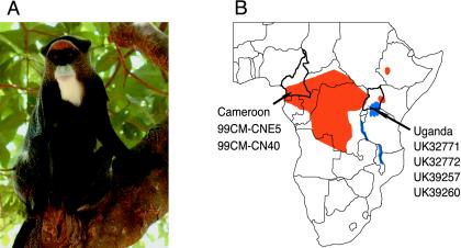
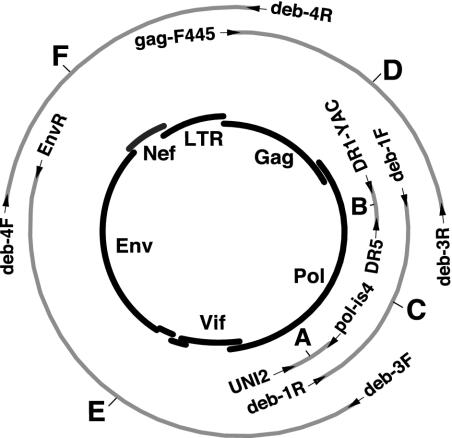
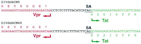
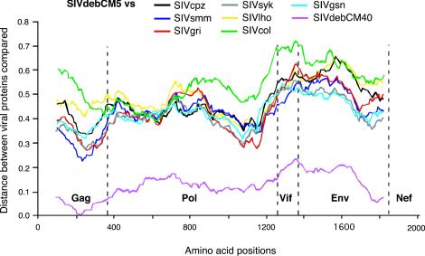
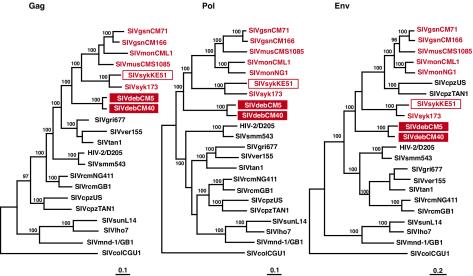
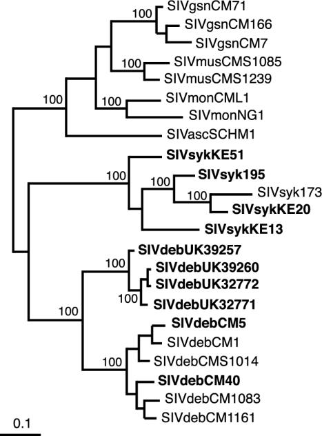
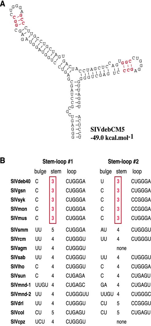
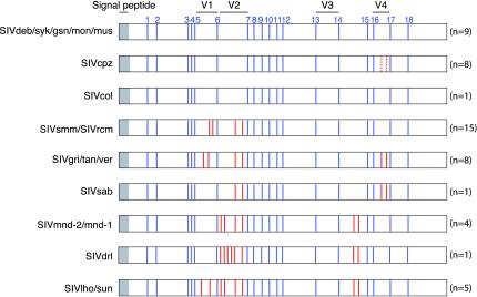
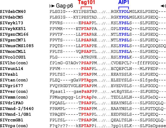
Nearly complete sequences of simian immunodeficiency viruses (SIVs) infecting 18 different nonhuman primate species in sub-Saharan Africa have now been reported; yet, our understanding of the origins, evolutionary history, and geographic distribution of these viruses still remains fragmentary. Here, we report the molecular characterization of a lentivirus (SIVdeb) naturally infecting De Brazza's monkeys (Cercopithecus neglectus). Complete SIVdeb genomes (9,158 and 9227 bp in length) were amplified from uncultured blood mononuclear cell DNA of two wild-caught De Brazza's monkeys from Cameroon. In addition, partial pol sequences (650 bp) were amplified from four offspring of De Brazza's monkeys originally caught in the wild in Uganda. Full-length (9068 bp) and partial pol (650 bp) SIVsyk sequences were also amplified from Sykes's monkeys (Cercopithecus albogularis) from Kenya. Analysis of these sequences identified a new SIV clade (SIVdeb), which differed from previously characterized SIVs at 40 to 50% of sites in Pol protein sequences. The viruses most closely related to SIVdeb were SIVsyk and members of the SIVgsn/SIVmus/SIVmon group of viruses infecting greater spot-nosed monkeys (Cercopithecus nictitans), mustached monkeys (Cercopithecus cephus), and mona monkeys (Cercopithecus mona), respectively. In phylogenetic trees of concatenated protein sequences, SIVdeb, SIVsyk, and SIVgsn/SIVmus/SIVmon clustered together, and this relationship was highly significant in all major coding regions. Members of this virus group also shared the same number of cysteine residues in their extracellular envelope glycoprotein and a high-affinity AIP1 binding site (YPD/SL) in their p6 Gag protein, as well as a unique transactivation response element in their viral long terminal repeat; however, SIVdeb and SIVsyk, unlike SIVgsn, SIVmon, and SIVmus, did not encode a vpu gene. These data indicate that De Brazza's monkeys are naturally infected with SIVdeb, that this infection is prevalent in different areas of the species' habitat, and that geographically diverse SIVdeb strains cluster in a single virus group. The consistent clustering of SIVdeb with SIVsyk and the SIVmon/SIVmus/SIVgsn group also suggests that these viruses have evolved from a common ancestor that likely infected a Cercopithecus host in the distant past. The vpu gene appears to have been acquired by a subset of these Cercopithecus viruses after the divergence of SIVdeb and SIVsyk.


Figures

FIG. 1.
FIG. 1.
Geographical distribution of De Brazza's monkeys (Cercopithecus neglectus) in sub-Saharan Africa. (A) Female De Brazza's monkey from Cameroon. (B) Geographic origin of the De Brazza's monkeys analyzed in this study. Animals 99CM-CNE5 and 99CM-CN40 were wild caught in Cameroon (60). UK32771, UK32772, UK3925, and UK39260 were captive born in safari parks in the United Kingdom, but are progeny of De Brazza's monkeys originally imported from Uganda. Cameroon and Uganda are shown in relation to the species' natural range (orange) in sub-Saharan Africa (country borders are shown in black).
FIG. 2.
FIG. 2.
Amplification of full-length SIVdeb and SIVsyk sequences from uncultured monkey PBMC DNA. The positions of the various amplification products are shown in relation to an unintegrated (single-LTR) circular intermediate of SIV (depicted in the center) with major genomic regions indicated. Primer sets (indicated by arrows) and fragment designations (A to F) are identical to those in Table 2.
FIG. 3.
FIG. 3.
Vpr-tat intergenic region in SIVdeb. SIVdeb vpr (red) and first exon of tat (green) genes are separated by a 19-bp noncoding sequence (black) in two different SIVdeb strains (CM5 and CM40). The deduced amino acid sequences of the C terminus of Vpr and the N terminus of Tat are indicated. SA indicates the splice acceptor site for the tat mRNA.
FIG. 4.
FIG. 4.
Diversity plots of concatenated protein sequences illustrating the extent of genetic difference between SIVdebCM5 and other primate lentiviruses. The proportion of amino acid sequence differences between SIVdebCM5 and other SIV strains are shown in different colors. Values are plotted at the midpoint of the sequence window on the x axis, with the N termini of Gag, Pol, Vif, Env, and Nef indicated. The y axis indicates the distance between the viral proteins (0.1 = 10% difference).
FIG. 5.
FIG. 5.
Phylogenetic relationship of SIVdeb strains to other primate lentiviruses. Midpoint-rooted maximum-likelihood trees were inferred from protein sequence alignments. The numbers on internal branches indicate estimated posterior probabilities (only values of 95% or greater are shown); the scale bars indicate 0.1 (Gag and Pol) and 0.2 (Env) substitutions per site. SIVs from Cercopithecus species (excluding the C. lhoesti group, whose taxonomic classification has been disputed [74]) are shown in red, including three new sequences obtained in this study (boxed).
FIG. 6.
FIG. 6.
Phylogenetic relationships of SIVdeb from Cameroon and Uganda and SIVsyk from Kenya to SIVs from other Cercopithecus species (excluding the C. lhoesti group). Maximum-likelihood trees were inferred from a partial (170-amino-acid) Pol protein sequence alignment and rooted as for Fig. 5. The numbers on internal branches indicate estimated posterior probabilities (only values of 95% or greater are shown); the scale bar indicates 0.1 substitution per site. Sequences obtained in this study are shown in bold.
FIG. 7.
FIG. 7.
Secondary structure predictions of SIV TAR elements. (A) The secondary structure for the SIVdebCM5 TAR with the lowest free energy value (−49.0 kcal/mol) is shown. The predicted SIVdebCM40 TAR secondary structure was almost identical, with a free energy value of −47.6 kcal/mol (not shown). (B) Comparison of TAR elements from other primate lentiviruses. The five Cercopithecus SIVs listed at the top (SIVdeb, SIVgsn, SIVsyk, SIVmon, and SIVmus) share a unique 3-bp stem separating the bulge and the terminal loop (highlighted in red).
FIG. 8.
FIG. 8.
Position of cysteine residues in the Env glycoprotein surface subunit of primate lentiviruses. The gp120 domain is depicted, with signal peptide and variable regions (V1-V4) indicated. Blue bars indicate the relative location of the 18 cysteine residues (numbered 1 to 18 at the top) that are conserved among all primate lentiviruses; red bars depict additional cysteine residues that are present in a subset of SIV strains (broken lines indicate one cysteine pair unique to SIVcpzCAM5). The number of strains per SIV group is indicated in parentheses.
FIG. 9.
FIG. 9.
Alignment of primate lentiviral Gag p6 protein sequences. Only N- and C-terminal sequences are shown, with strain designations indicated on the left. For SIVlho, SIVtan, SIVver, SIVsmm, and SIVcpz, consensus sequences (con) are shown (45). Uppercase letters indicate amino acids that are conserved in all sequences. Lowercase letters (or a question mark [?]) indicate amino acids conserved in more (or fewer) than 50% of sequences used to generate the consensus sequence. Dashes indicate spaces introduced to optimize the alignment. Tsg101 (PT/SAP) and AIP1 (YPD/SL) binding motifs are shown in red and blue, respectively.

Similar articles

Cited by

References

    1. Allan, J. S., M. Short, M. E. Taylor, S. Su, V. M. Hirsch, P. R. Johnson, G. M. Shaw, and B. H. Hahn. 1991. Species-specific diversity among simian immunodeficiency viruses from African green monkeys. J. Virol. 65:2816-2828. - PMC - PubMed
    1. Bailes, E., R. R. Chaudhuri, M. L. Santiago, F. Bibollet-Ruche, B. H. Hahn, and P. M. Sharp. 2002. The evolution of primate lentiviruses and the origins of AIDS, p. 65-96. In T. A. Leitner (ed.), The molecular epidemiology of human viruses. Kluwer Academic Publishers, Boston, Mass.
    1. Bailes, E., F. Gao, F. Bibollet-Ruche, V. Courgnaud, M. Peeters, P. A. Marx, B. H. Hahn, and P. M. Sharp. 2003. Hybrid origin of SIV in chimpanzees. Science 300:1713.. - PubMed
    1. Barlow, K. L., A. O. Ajao, and J. P. Clewley. 2003. Characterization of a novel simian immunodeficiency virus (SIVmonNG1) genome sequence from a mona monkey (Cercopithecus mona). J. Virol. 77:6879-6888. - PMC - PubMed
    1. Beer, B. E., E. Bailes, G. Dapolito, B. J. Campbell, R. M. Goeken, M. K. Axthelm, P. D. Markham, J. Bernard, D. Zagury, G. Franchini, P. M. Sharp, and V. M. Hirsch. 2000. Patterns of genomic sequence diversity among their simian immunodeficiency viruses suggest that L'Hoest monkeys (Cercopithecus lhoesti) are a natural lentivirus reservoir. J. Virol. 74:3892-3898. - PMC - PubMed

Publication types

MeSH terms