The 3' cis-acting genomic replication element of the severe acute respiratory syndrome coronavirus can function in the murine coronavirus genome
- PMID: 15220462
- PMCID: PMC434098
- DOI: 10.1128/JVI.78.14.7846-7851.2004
The 3' cis-acting genomic replication element of the severe acute respiratory syndrome coronavirus can function in the murine coronavirus genome
Abstract
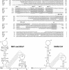
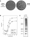
The 3' untranslated region (3' UTR) of the genome of the severe acute respiratory syndrome coronavirus can functionally replace its counterpart in the prototype group 2 coronavirus mouse hepatitis virus (MHV). By contrast, the 3' UTRs of representative group 1 or group 3 coronaviruses cannot operate as substitutes for the MHV 3' UTR.
Figures


Similar articles
-
Common RNA replication signals exist among group 2 coronaviruses: evidence for in vivo recombination between animal and human coronavirus molecules.Virology. 2003 Oct 10;315(1):174-83. doi: 10.1016/s0042-6822(03)00511-7. Virology. 2003. PMID: 14592769 Free PMC article.
-
Characterization of the RNA components of a putative molecular switch in the 3' untranslated region of the murine coronavirus genome.J Virol. 2004 Jan;78(2):669-82. doi: 10.1128/jvi.78.2.669-682.2004. J Virol. 2004. PMID: 14694098 Free PMC article.
-
Stem-loop 1 in the 5' UTR of the SARS coronavirus can substitute for its counterpart in mouse hepatitis virus.Adv Exp Med Biol. 2006;581:105-8. doi: 10.1007/978-0-387-33012-9_18. Adv Exp Med Biol. 2006. PMID: 17037514 Free PMC article. No abstract available.
-
Development of mouse hepatitis virus and SARS-CoV infectious cDNA constructs.Curr Top Microbiol Immunol. 2005;287:229-52. doi: 10.1007/3-540-26765-4_8. Curr Top Microbiol Immunol. 2005. PMID: 15609514 Free PMC article. Review.
-
SARS: lessons learned from other coronaviruses.Viral Immunol. 2003;16(4):461-74. doi: 10.1089/088282403771926292. Viral Immunol. 2003. PMID: 14733734 Review.
Cited by
-
Regulation viral RNA transcription and replication by higher-order RNA structures within the nsp1 coding region of MERS coronavirus.Sci Rep. 2024 Aug 23;14(1):19594. doi: 10.1038/s41598-024-70601-5. Sci Rep. 2024. PMID: 39179600 Free PMC article.
-
A three-stemmed mRNA pseudoknot in the SARS coronavirus frameshift signal.PLoS Biol. 2005 Jun;3(6):e172. doi: 10.1371/journal.pbio.0030172. Epub 2005 May 17. PLoS Biol. 2005. PMID: 15884978 Free PMC article.
-
Structural and functional conservation of cis-acting RNA elements in coronavirus 5'-terminal genome regions.Virology. 2018 Apr;517:44-55. doi: 10.1016/j.virol.2017.11.025. Epub 2017 Dec 6. Virology. 2018. PMID: 29223446 Free PMC article.
-
Coronavirus cis-Acting RNA Elements.Adv Virus Res. 2016;96:127-163. doi: 10.1016/bs.aivir.2016.08.007. Epub 2016 Sep 6. Adv Virus Res. 2016. PMID: 27712622 Free PMC article. Review.
-
Analysis of Emerging Variants in Structured Regions of the SARS-CoV-2 Genome.Evol Bioinform Online. 2021 May 5;17:11769343211014167. doi: 10.1177/11769343211014167. eCollection 2021. Evol Bioinform Online. 2021. PMID: 34017166 Free PMC article.
References
Publication types
MeSH terms
Substances
Grants and funding
LinkOut - more resources
Full Text Sources
Other Literature Sources

