. 2004 Jul 6;101(27):10211-6.
doi: 10.1073/pnas.0403709101. Epub 2004 Jun 25.

A probable Na+(K+)/H+ exchanger on the chloroplast envelope functions in pH homeostasis and chloroplast development in Arabidopsis thaliana

Affiliations

A probable Na+(K+)/H+ exchanger on the chloroplast envelope functions in pH homeostasis and chloroplast development in Arabidopsis thaliana

Chun-Peng Song et al. Proc Natl Acad Sci U S A. .

Abstract

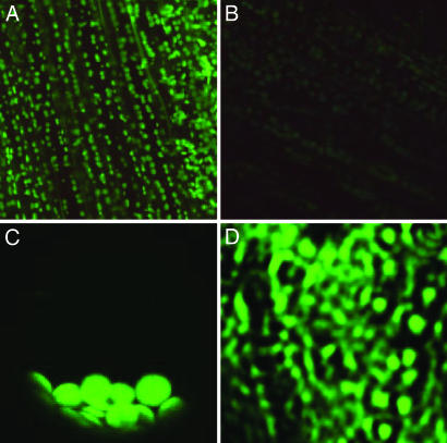
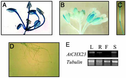
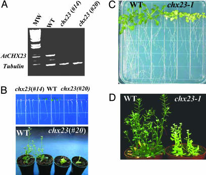
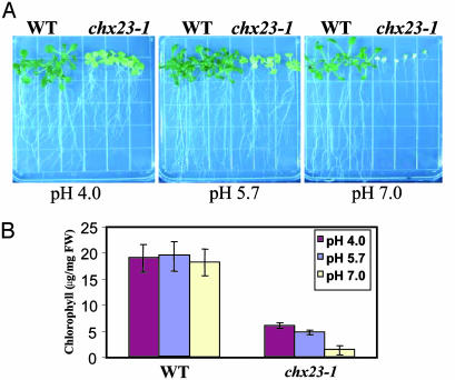
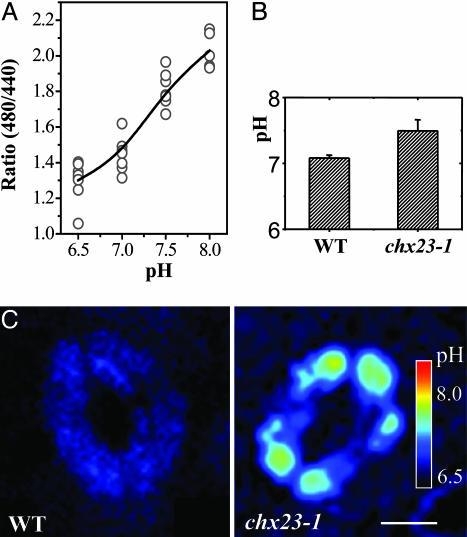
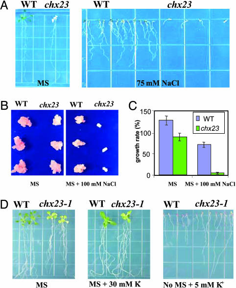
Electroneutral monovalent cation/proton antiport across the chloroplast envelope has been shown previously to have an important regulatory effect on stromal pH and thereby on photosynthetic carbon reduction. Here we report that an Arabidopsis nuclear gene, AtCHX23, encodes a putative Na(+)(K(+))/H(+) exchanger and functions in the adjustment of pH in the cytosol and possibly in maintaining a high pH level in the chloroplast stroma. The AtCHX23 protein is localized in the chloroplast envelope. Plastids from chx23 mutants had straight thylakoids but lacked grana lamellae. chx23 mutant leaves were pale yellow and had a much reduced chlorophyll content. The chlorophyll content of chx23 was increased by growing in medium at low (4.0) pH and decreased by growing at high (7.0) pH. The cytosolic pH in the leaves of the mutant was significantly higher than that in the wild type. chx23 mutants displayed a high sensitivity to NaCl. Together, these data indicate that CHX23 is a probable chloroplast Na(+)(K(+))/H(+) exchanger important for pH homeostasis and chloroplast development and function.


Figures

Fig. 1.
Fig. 1.
Subcellular localization of the AtCHX23-GFP fusion protein. (A) Chloroplast localization of AtCHX23-GFP in cells from main vein and leaves. (B) Autofluorescence of leaf cells under same conditions as in A. (C) A single protoplast isolated from the transgenic lines with pEZTL-CHX23-GFP. The protoplast had been centrifuged, so the chloroplasts are bunched together at the bottom. (D) The chloroplast envelope membrane vesicles purified from AtCHX23-GFP transgenic plants.
Fig. 2.
Fig. 2.
Histochemical localization of GUS activity directed by AtCHX23 promoter::GUS fusions in transgenic Arabidopsis. (A) Transgenic leaves carrying pAtCHX23-GUS. (B) Transgenic flowers carrying pAtCHX23-GUS. (C) Silique. (D) Transgenic roots carrying pAtCHX23-GUS. (E) RT-PCR shows the expression of chx23 in different tissues. L, leaves; R, roots; F, flowers; S, stems.
Fig. 3.
Fig. 3.
Phenotype of chx23 mutants. (A) AtCHX23 gene silencing by double-stranded RNAi. RT-PCR with wild type and two independent lines of chx23 using chx23-specific primers. Tubulin primers were used in PCR as an internal control. (B) The seeds of Arabidopsis wild type and chx23 (#14 and #20) were germinated in MS agar medium. The pictures were taken 7 and 20 d after germination and show the phenotype of chx23 at different developmental stages. (C) chx23-1 mutant from TILLING was germinated in MS for 15 d. (D) chx23-1 grown in soil for 25 d.
Fig. 4.
Fig. 4.
Ultrastructure of chloroplasts from wild type and the chx23-1 mutant. All pictures are transmission electron microscope images from ultrathin sections from leaves of 14-d-old seedlings. (Bars = 1 μm.)
Fig. 5.
Fig. 5.
Effect of pH on chx23 mutant growth and chlorophyll synthesis. Arabidopsis seeds were germinated in MS agar medium at different pH levels (4.0, 5.7, and 7.0). (A) Effects of different pH levels on the growth of wild type and chx23-1. The pictures were taken 20 d after germination. (B) Effects of different pH levels on the chlorophyll content from wild type and chx23 mutants. Chlorophyll was extracted by using N′-dimethylformamide and determined photometrically according to described procedures (29). Presented values are the means from six measurements of six seedlings (15 d) ± SD.
Fig. 6.
Fig. 6.
Cytoplasmic pH in wild-type and chx23 plants. (A) In situ calibration was performed by using 10 μg/ml nigericin and 100 mM KCl in buffers of different pH. Pixel values were averaged over recticular regions (15–30 μm2) manually located on each wavelength image. A single ratio value was calculated from the average 488 intensity divided by the average 442 intensity for this region, after subtraction of the corresponding image backgrounds. Values >200 were masked in both images to exclude pixels approaching saturation. (B) The pH changes in guard cells of wild type and chx23-1. The conversion from ratio values to pH derived from the in vivo calibration is indicated as the average cytosolic pH (mean ± SD, n = 8 cells). (C) pH distribution of guard cells was monitored by pseudoratiometric confocal imaging of the fluorescence from the pH-sensitive dye BCECF-AM through dual-excitation confocal ratio measurement. The pseudocolor bar in C represents a pH range from ≈8.0 to 6.5. (Bar = 10 μm.)
Fig. 7.
Fig. 7.
The effects of NaCl and KCl on the phenotype of chx23 mutants. (A) The germination of seeds from wild type and chx23 mutants. The pictures were taken 12 d after germination in MS agar plates without or with 75 mM NaCl. (B) Effects of NaCl on relative growth rate of wild type and chx23 mutant calli. Callus was generated from seedlings of the wild type and mutants, by incubation on callus-initiating medium containing MS salts, 2% sucrose, 3 mg/liter 2,4-D, 50 μg/liter and 0.8% agar for 2 weeks. (C) Effects of NaCl on relative growth rate of Arabidopsis calli. Calli were incubated in the corresponding medium for 24 d. Data are the means ± SD of three independent experiments with three replicates per experiment. (D) Wild-type and chx23 plants grown in MS medium or MS supplemented with 30 mM KCl or no MS salts supplemented with 5 mM KCl.

References

    1. Mitchell, P. (1961) Nature 191, 144-148. - PubMed
    1. Jagendorf, A. T. & Uribe, A. T. (1966) Proc. Natl. Acad. Sci. USA 55, 170-177. - PMC - PubMed
    1. Werdan, K., Heldt, H. W. & Milovancev, M. (1975) Biochim. Biophys. Acta 396, 276-292. - PubMed
    1. Heldt, H. W., Wedan, K. W., Milovancev, M. & Geller, G. (1973) Biochim. Biophys. Acta 314, 224-241. - PubMed
    1. Felle, H. H. (2001) Plant Biol. 3, 577-591.

Publication types

MeSH terms