. 2004 Jul 6;101(27):10000-5.
doi: 10.1073/pnas.0402784101. Epub 2004 Jun 25.

Activity-based probes for the proteomic profiling of metalloproteases

Affiliations

Activity-based probes for the proteomic profiling of metalloproteases

Alan Saghatelian et al. Proc Natl Acad Sci U S A. .

Abstract

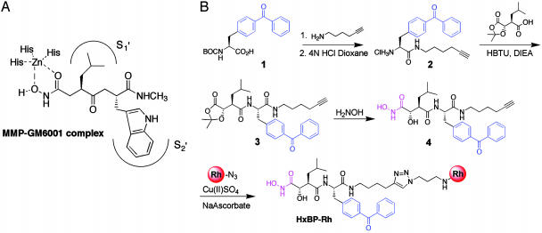
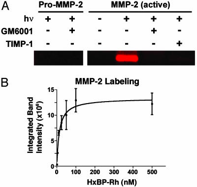
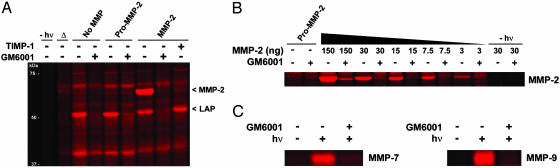
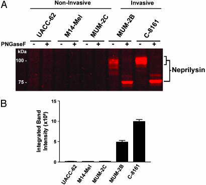
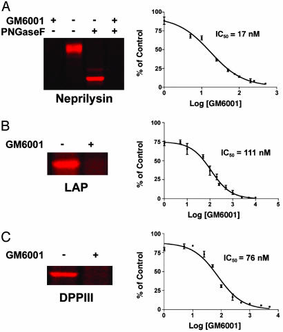
Metalloproteases (MPs) are a large and diverse class of enzymes implicated in numerous physiological and pathological processes, including tissue remodeling, peptide hormone processing, and cancer. MPs are tightly regulated by multiple posttranslational mechanisms in vivo, hindering their functional analysis by conventional genomic and proteomic methods. Here we describe a general strategy for creating activity-based proteomic probes for MPs by coupling a zinc-chelating hydroxamate to a benzophenone photocrosslinker, which promote selective binding and modification of MP active sites, respectively. These probes labeled active MPs but not their zymogen or inhibitor-bound counterparts and were used to identify members of this enzyme class up-regulated in invasive cancer cells and to evaluate the selectivity of MP inhibitors in whole proteomes. Interestingly, the matrix metalloproteinase inhibitor GM6001 (ilomastat), which is currently in clinical development, was found to also target the neprilysin, aminopeptidase, and dipeptidylpeptidase clans of MPs. These results demonstrate that MPs can display overlapping inhibitor sensitivities despite lacking sequence homology and stress the need to evaluate MP inhibitors broadly across this enzyme class to develop agents with suitable target selectivities in vivo. Activity-based profiling offers a powerful means for conducting such screens, as this approach can be carried out directly in whole proteomes, thereby facilitating the discovery of disease-associated MPs concurrently with inhibitors that selectively target these proteins.


Figures

Fig. 1.
Fig. 1.
Design and synthesis of an MP-directed activity-based probe, HxBP-Rh. (A) General interactions between a broad-spectrum reversible hydroxamate inhibitor (GM6001) and MMP active sites based on a combination of structure-activity (45) and crystallographic data (51). (B) Synthesis of HxBP-Rh for activity-based profiling of MPs. Hydroxamate, benzophenone, and rhodamine groups are shown in magenta, blue, and red, respectively. Details regarding the synthesis and characterization of HxBP-Rh are provided in the supporting information. HBTU, 2-(1H-benzotriazole-1-yl)-1,1,3,3-tetramethyluronium hexafluorophosphate; DIEA, diisopropylethylamine.
Fig. 2.
Fig. 2.
Activity-based labeling of purified MMP-2 by HxBP-Rh. (A) HxBP-Rh (100 nM) was incubated with purified samples of pro-MMP-2 (30 ng) or MMP-2 (30 ng) with or without inhibitors [GM6001 (5 μM) or TIMP-1 (80 ng)] for 15 min before photocrosslinking by exposure to UV light (hν). Samples were then analyzed by SDS/PAGE and in-gel fluorescence scanning. HxBP-Rh labeled active MMP-2 but not pro-MMP-2 or inhibitor-bound MMP-2. (B) Concentration dependence of MMP-2 labeling by HxBP-Rh. Labeling of MMP-2 was saturated at ≈100 nM HxBP-Rh. Labeling was measured by in-gel fluorescence scanning (integrated band intensities are given in arbitrary units). Each data point corresponds to the average of two independent trials.
Fig. 3.
Fig. 3.
Activity-based labeling of MMPs in whole proteomes by HxBP-Rh. (A) Purified MMP-2 (100 ng) or pro-MMP-2 (100 ng) was added to a mouse kidney proteome (15 μl, 1 μg of protein per microliter) and treated with HxBP-Rh (100 nM) for 15 min before photocrosslinking and analysis by SDS/PAGE and in-gel fluorescence scanning. Only MMP-2 was labeled by HxBP-Rh, and this labeling was blocked by GM6001 (5 μM) and TIMP-1 (200 ng). No protein labeling was observed in the absence of exposure to UV light. Δ, heat-denatured proteome (containing 100 ng of MMP-2). Also highlighted in this profile is an endogenous GM6001-sensitive enzyme activity labeled by HxBP-Rh, which was identified by using a trifunctional HxBP probe as LAP (see Fig. 5B). (B) HxBP-Rh labeling of a serial dilution of purified MMP-2 added to a mouse kidney proteome. HxBP-Rh could detect as few as 3 ng of active MMP-2 (corresponding to 3 nM enzyme in a background of 15 μlof1 μg/μl proteome). HxBP-Rh did not label pro-MMP-2 (150 ng). (C) HxBP-Rh labeling of MMP-7 and MMP-9 in proteomes. MMP-7 and MMP-9 (30 ng) were added to the mouse kidney proteome (15 μl, 1 μg/μl), and the samples were treated with HxBP-Rh (100 nM) and analyzed as described above. HxBP-Rh labeled active, but not GM6001-inhibited, MMP-7 and MMP-9.
Fig. 4.
Fig. 4.
HxBP-Rh identifies neprilysin as an MP activity dramatically upregulated in invasive human melanoma cell lines. (A) HxBP-Rh labeling profiles of membrane proteomes from a panel of human melanoma cell lines. An HxBP-Rh-labeled glycoprotein highly up-regulated in invasive melanoma lines (MUM-2B and C-8161) compared with noninvasive melanoma lines (UACC-62, M14-Mel, and MUM-2C) was identified as neprilysin by using a trifunctional HxBP probe (see Methods for more details). Deglycoslyation was accomplished by treating a portion of each HxBP-Rh-labeled proteome with PNGaseF before analysis. (B) Quantitation of neprilysin activity in melanoma membrane proteomes by in-gel fluorescence scanning (n = 3 per group).
Fig. 5.
Fig. 5.
HxBP-Rh identifies several MPs outside the MMP family that are inhibited by GM6001, including neprilysin (A), LAP (B), and DPPIII (C). (Left) Shown is representative labeling of MPs in whole proteomes by HxBP-Rh (100 nM) and inhibition by GM6001 (5 μM). Note that PNGaseF lanes are not shown for LAP and DPPIII because treatment with this glycosidase did not alter the migration of these MPs by SDS/PAGE. Neprilysin was identified in the secreted proteome of invasive human melanoma cell lines (see Fig. 4), whereas LAP and DPPIII were identified in soluble proteomes from mouse kidney (see Fig. 3A) and the human breast cancer cell line MCF7, respectively (for a full profile of HxBP-Rh labeling of the MCF7 soluble proteome, see Fig. 6, which is published as supporting information on the PNAS web site). (Right) Shown is the concentration-dependence of inhibition of HxBP-Rh labeling by GM6001 (each data point corresponds to the average of three independent trials and is presented as a percentage of control reactions conducted without GM6001). From these curves, IC50 values of 17 nM (12–23 nM, 95% confidence limits), 111 nM (83–149 nM, 95% confidence limits), and 76 nM (53–109 nM, 95% confidence limits), were calculated for the inhibition of neprilysin, LAP, and DPPIII, respectively, by GM6001.

Similar articles

Cited by

References

    1. Patterson, S. D. & Aebersold, R. (2003) Nat. Genet. 33, 311-323. - PubMed
    1. Adam, G. C., Sorensen, E. J. & Cravatt, B. F. (2002) Mol. Cell. Proteomics 1, 781-790. - PubMed
    1. Michnick, S. W. (2004) Drug Discovery Today 9, 262-267. - PubMed
    1. Patton, W. F., Schulenberg, B. & Steinberg, T. H. (2002) Curr. Opin. Biotechnol. 13, 321-328. - PubMed
    1. Gygi, S. P., Rist, B., Gerber, S. A., Turecek, F., Gelb, M. H. & Aebersold, R. (1999) Nat. Biotechnol. 17, 994-999. - PubMed

Publication types

Substances